Heath's practical anatomy : a manual of dissections / [Christopher Heath].
- Christopher Heath
- Date:
- 1893
Licence: Public Domain Mark
Credit: Heath's practical anatomy : a manual of dissections / [Christopher Heath]. Source: Wellcome Collection.
65/800 page 43
![The Supra-scapular Nerve (Fig. 15, i) (from the anterior branches of the 5th and 6th cervical nerves) gives a branch to the shoulder-joint and passes through the supra-scapular notch and beneath the ligament, to be distributed to the supra- and infra- spinatus muscles. The Omo-hyoid Muscle arises from about half an inch of the superior border of the scapula, immediately behind the supra- scapular notch, and from the transverse or posterior ligament over it. Its relations and insertian are seen in the dissection of the neck. [The limb is to be turned over and the humerus rotated outwards to put the fibres of the subscapularis on the stretch ; they should then be cleaned, care being taken of the subscapular nerves entering the muscle. The axillary vessels and nerves should be tied to the coracoid process, so as to bring them as nearly as may be into their proper positions.] The Subscapularis Muscle (Fig. 9, lo) is covered by a thin fascia, and has been already seen to form j^art of the posterior wall of the axilla. It arises from the whole of the venter scapulae, except the part to which the serratus magnus is attached and the portion nearest the neck of the bone, and has several tendinous septa be- tween its fibres, which are attached to the ridges on the surface of the bone, and bear some resemblance to those of the deltoid. It is inserted into the lesser tuberosity of the humerus and the bone below it for an inch, and into the capsular ligament of the shoulder-joint, a large bursa intervening between the muscle and the neck of the scapula, which almost always communicates with the cavity of the joint over the upper border of the tendon. The subscapularis is an internal rotator and adductor of the humerus, and is supiolied by the short subscapular nerves. An important action of the supra- and infra-spinati, teres minor, and subscapularis muscles is their combination to keep the head of the humerus in its proper relation to the glenoid cavity, and thus prevent dislocation. In the post-mortem relaxed condition of the parts, the head of the humerus can be drawn away from the glenoid cavity to the full extent of the loose capsular ligament. The Subscapular Werves (Fig. 7) are branches of the posterior cord of the brachial plexus, and are three in number. The long or middle subscapular nerve has been already seen in the axilla, and can still be traced to the latissimus dorsi; the short subscapular nerves (upper and lower) are now to be followed, the upper to the subscapularis muscle, the lower to the subscapularis and teres major.](https://iiif.wellcomecollection.org/image/b20417457_0065.jp2/full/800%2C/0/default.jpg)
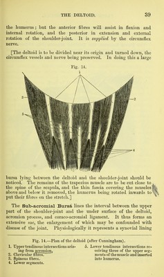


 No text description is available for this image
 No text description is available for this image
 No text description is available for this image