Heath's practical anatomy : a manual of dissections / [Christopher Heath].
- Christopher Heath
- Date:
- 1893
Licence: Public Domain Mark
Credit: Heath's practical anatomy : a manual of dissections / [Christopher Heath]. Source: Wellcome Collection.
66/800 page 44
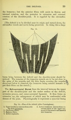
![The Subscapular Artery (p. 19) is still to be seen running along the axillary border of the scapula, ^d its branches should The Infra-scapular Artery is deriveti from the dorsal branch of the subscapular artery. It passes into the venter scajjulse, beneath the subscapularis muscle, which must be divided to expose it, and anastomoses with the neighbouring vessels. The Posterior Scapular Artery is to be found between the serratus magnus and the rhomboidei, and its anastomoses upon the dorsal and ventral surfaces of the scapula should be defined. By removing the muscular fibres from both surfaces of the scapula a very abundant network of vessels will be seen, formed by anasto- mosing branches from the arteries which have been already traced to the scapula; viz., the subscapular with its dorsal branch [axillary], the supra-scapular [thyroid axis], and the posterior scapular [thyroid axis or subclavian]. [The fore-arm and hand are to be doubled under the upper arm, which is to be placed on the table with the back upwards, and the scapula is to be drawn down with hooks so as to put the triceps on the stretch. When the skin has been removed from the back of the arm, two external cutaneous branches of the musculo-spiral nerve should be noticed.] Cutaneous Nerves.—The wpper external cutaneous branch of the musculo-spiral (Fig. 8, 7) appears about the middle of the outer side of the arm, and runs downwards and forwards along the cephalic vein to the upper part of the fore-arm ; the lower external, of larger size (Fig. 16, 6), appears close above the external condyle, and will be afterwards traced down the back of the fore- arm to the wrist; and the third or internal cutaneous branch pierces the fascia near the tendon of the teres major and supplies an area on the inner side of the arm behind that of the intercosto- humeral nerve (Fig. 8, 6). Branches from the internal cutaneous and lesser internal cutaneous nerves will be found on the inner side of the limb, and filaments of the circumflex nerve run downwards over the back of the arm, and upwards over the lower part of the deltoid (Fig. 16). [When the strong deep fascia of the arm has been divided, the fibres of the triceps muscle should be cleaned, and a large bursa between the triangular posterior surface of the ulna and the skin should be noticed.] be thoroughly followed out. The Back of the Arm.](https://iiif.wellcomecollection.org/image/b20417457_0066.jp2/full/800%2C/0/default.jpg)


 No text description is available for this image
 No text description is available for this image
 No text description is available for this image