Descriptive catalogue of the pathological specimens contained in the museum of the Royal College of Surgeons of England.
- Bader, Charles.
- Date:
- 1864
Licence: Public Domain Mark
Credit: Descriptive catalogue of the pathological specimens contained in the museum of the Royal College of Surgeons of England. Source: Wellcome Collection.
Provider: This material has been provided by UCL Library Services. The original may be consulted at UCL (University College London)
52/106
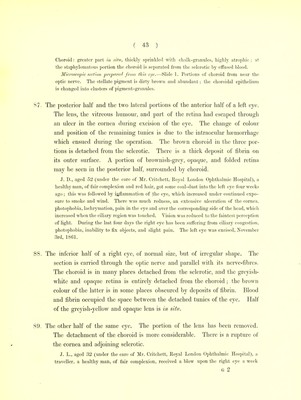
![opaque ; the latter is detached from the choroid, and may be traced from the optic nerve to where it disappears behind the grey-brown substance. The choroid has a deep-brown colour ; it is in most places slightly detached from the sclerotic by clots similar to the one in the vitreous space. A large rent is observed across the cornea, near its margin. No lens was found. 95. The other half of the same eye. The adhesion of the anterior surface of the iris, the margins of the rent in the cornea, and the change of colour in the optic nerve may be observed. E. W., aged 23 (under the care of Mr. Poland, Royal London Ophthalmic Hospital), a healthy woman, of dark complexion, received a blow on the right eye twelve days ago. Vision was lost at once ; much blood escaped from the eye. The fellow eye began to sym- pathize at once, as evinced by photophobia, inability to fix the eye on objects, and pain on touching the ciliary region. The right eye was excised, June 22nd, 1861. Dissection.—Sbape and size normal. Cornea transparent; a rupture extended through the tunics of the ciliary region, at its lowest margin. 96. The lateral half of a considerably shrunken left eye, the section being carried through the optic nerve, and parallel with its nerve-fibres. The thickened sclerotic is in apposition with the dirty-brown choroid ; it is drawn inwards in its superior and inferior equatorial regions. The place of the vitreous and retina is occupied by dense white fibrous tissue. 97. The other half of the same eye. J. S., aged 57 (under the care of Mr. Poland, Royal London Ophthalmic Hospital), a healthy man, of fair complexion, five months ago had a cataract removed from the left eye by Shuft's method (iridectomy and extraction of the cataract with a large scoop). Much straining and coughing followed, the night after the operation ] the eye finally suppurated. The sympathy of the other eye, and the great pain in the shrinking, made excision necessary. The left eye was excised, October 12th, 1861. 98. A lateral portion of a considerably shrunken and irregularly shaped left eye, the section being carried through the middle of the cornea. No retina, vitreous, or lens was found. The posterior part of the sclerotic is much thickened j within it a part of the choroid may be seen. M. L. L., aged 17 (under the care of Mr. Poland, Royal London Ophthalmic Hospital), a](https://iiif.wellcomecollection.org/image/b21641857_0054.jp2/full/800%2C/0/default.jpg)