The surgical diseases of the ear / by Prof. von Troltsch. The mechanism of the ossicles and the membrana tympani / by Prof. Helmholtz ; translated from the German by James Hinton.
- Tröltsch, Anton Friedrich, Freiherr von, 1829-1890.
- Date:
- 1874
Licence: Public Domain Mark
Credit: The surgical diseases of the ear / by Prof. von Troltsch. The mechanism of the ossicles and the membrana tympani / by Prof. Helmholtz ; translated from the German by James Hinton. Source: Wellcome Collection.
Provider: This material has been provided by Royal College of Physicians, London. The original may be consulted at Royal College of Physicians, London.
137/180 (page 121)
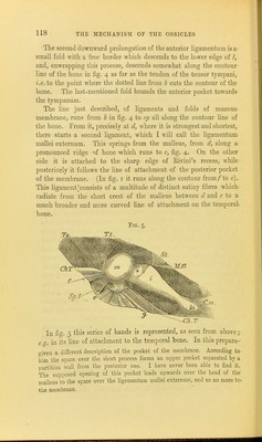
![The tightness of these ligaments is, under natural conditions, still further increased by the elastic tension of the relatively strong muscle, the tensor tympani, whose tendon is inserted at the begin- ning of the handle of the malleus, on the anterior half of its surface, which is turned mediurawards and faces the tube, a little lower down than where the short process projects from its side at the u])per part of the handle^ a little below the short process. Eig. 9 shows the somewhat oblique line of attachment of the tendon running from above downwards and backwards. The muscle lies, as we know, in a peculiar canal of bone, which runs over the Eustachian tube, through which the tympanum com- municates with the throat. The further end of the muscle arises outside this canal from the under surfj^ce of the pyramidal portion of the petrous bone, and from the cartilaginous portion of the Eustachian tube. Thence it proceeds through its canal, the end of ■which, opening on the tympanum, forms a hook-shaped prominence, round which the tendon winds, so as to run finally across the tym- panum to the malleus. The direction of the tendon is nearly per- pendicular to the plane in which the edge of the membrane lies, so that its Hue of action only deviates from it slightly downwards and forwards. On the other hand, it forms a pretty acute angle with tlie lower part of the handle of the malleus, and with the anterior portion of its axis of rotation. The tensor tympani is a feathered muscle; it springs from the periosteum of the upper surface of the bony canal in which it lies. Its tendon lies on its under side, and turns a smooth free surface towards the smooth ])eriosteum. The muscle-fibres are pretty short, and the tendon, therefore, contracts as far as the lower end of the canal. The tube of the periosteum which encloses the muscle is prolonged also over the free portion of the tendon that runs through the tympanum, externally covered by Its mucous membrane. Toynbee gives the name of tensor ligament of the membrane to this sheath of the free portion of the tendon. The isolation of the tendon and its sheath from one another appears to be more or less perfect when one compares different accounts. I myself have found, in a preparation in the anatomical collection at Bonn, a perfectly smooth and free tendon inside the sheath, as ioynbee describes. On the contrary, Henle has seen in microscopic sections the tendon and sheath blended together by moderately strong con- nective tissue. In consequence of the very small space for move-](https://iiif.wellcomecollection.org/image/b23984399_0137.jp2/full/800%2C/0/default.jpg)