Text-book of botany : morphological and physiological / by Julius Sachs ; translated and annotated by Alfred W. Bennett ; assisted by W.T. Thiselton Dyer.
- Date:
- 1875
Licence: Public Domain Mark
Credit: Text-book of botany : morphological and physiological / by Julius Sachs ; translated and annotated by Alfred W. Bennett ; assisted by W.T. Thiselton Dyer. Source: Wellcome Collection.
Provider: This material has been provided by the Royal College of Physicians of Edinburgh. The original may be consulted at the Royal College of Physicians of Edinburgh.
43/880 page 27
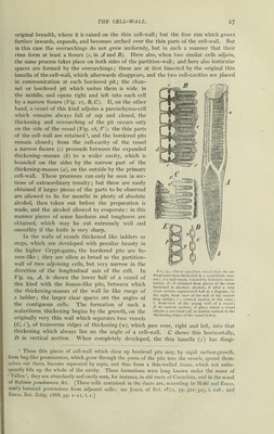
![original breadth, where it is raised on the thin cell-wall; but the free rim which grows further inwards, expands, and becomes arched over the thin parts of the cell-wall. But in this case the overarchings do not grow uniformly, but in such a manner that their rims form at least a fissure (c, in A and B). Here also, when two similar cells adjoin, the same process takes place on both sides of the partition-wall; and here also lenticular spaces are formed by the overarchings; these are at first bisected by the original thin lamella of the cell-wall, which afterwards disappears, and the two cell-cavities are placed in communication at each bordered pit; the chan- nel or bordered pit which unites them is wide in the middle, and opens rjght and left into each cell by a narrow fissure (Fig. 27, B, C). If, on the other hand, a vessel of this kind adjoins a parenchyma-cell which remains always full of sap and closed, the thickening and overarching of the pit occurs only on the side of the vessel (Fig. 28, V); the thin parts of the cell-wall are retained ■, and the bordered pits remain closed; from the cell-cavity of the vessel a narrow fissure (r) proceeds between the expanded thickening-masses (b) to a wider cavity, which is bounded on the sides by the narrow part of the thickening-masses (a), on the outside by the primary cell-wall. These processes can only be seen in sec- tions of extraordinary tenuity; but these are easily obtained if larger pieces of the parts to be observed are allowed to lie for months' in plenty of absolute alcohol, then taken out before the preparation is made, and the alcohol allowed to evaporate: in this manner pieces of some hardness and toughness are obtained, which may be cut extremely well and smoothly if the knife is very sharp. In the walls of vessels thickened like ladders 01- steps, which are developed with peculiar beauty in the higher Cryptogams, the bordered pits are fis- sure-like ; they are often as broad as the partition- wall of two adjoining cells, but very narrow in the direction of the longitudinal axis of the cell. In Fig. 29, A, is shown the lower half of a vessel of this kind with the fissure-like pits, between which the thickening-masses of the wall lie like rungs of a ladder; the larger clear spaces are the angles of the contiguous cells. The formation of such a scalariform thickening begins by the growth, on the originally very thin wall which separates two vessels (C, /), of transverse ridges of thickening (v), which pass over, right and left, into that thickening which always lies on the angle of a cell-wall. C shows this horizontally, D in vertical section. A\ hen completely developed, the thin lamella (/) has disap- 1 These thin pieces of cell-wall which close up bordered pits may, by rapid surface-growth, form bag-like prominences, which grow through the pores of the pits into the vessels, spread them- selves out there, become separated by septa, and thus form a thin-walled tissue, which not unfre- quently fills up the whole of the cavity. These formations were long known under the name of 1 alien ; they are abundantly and easily seen, for instance, in old roots of Cucurbita, and in the wood of Rtjbinia psendacacia, &c. [These cells contained in the ducts are, according to Mohl and Reess, really hernioid protrusions from adjacent cells; see Journ. of Hot. 1872, pp. 321-323, t. 126; and Reess, Bot. Zcitg. 1868, pp. 1-11, 1.1.] Fig. 29.—Pteris aquilina, vessel from the un- derground stem thickened in a scalariform man- ner; A a half-vessel, isolated by Schulze’s mace- ration ; B—D obtained from pieces of the stem hardened in absolute alcohol; B after a very clean section, represented half as a diagram ; to the right, front view of the wall of the vessels from within; c c vertical section of the same ; C front-view of the young wall of a vessel; D its vertical section; E place where a vessel adjoins a succulent cell, in section vertical to the thickening-ridges of the vessel (X800).](https://iiif.wellcomecollection.org/image/b21981437_0043.jp2/full/800%2C/0/default.jpg)