The cultivation of human tumour tissue in vitro : preliminary note / by David Thomson and John Gordon Thomson.
- Thomson, D. (David), 1884-
- Date:
- 1914]
Licence: Public Domain Mark
Credit: The cultivation of human tumour tissue in vitro : preliminary note / by David Thomson and John Gordon Thomson. Source: Wellcome Collection.
3/8
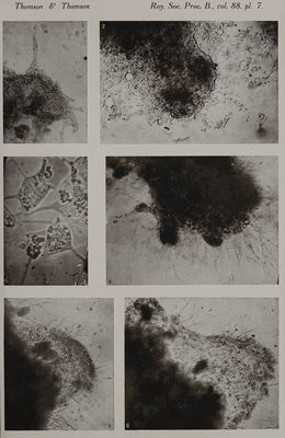
![[Reprinted from the PROCEEDINGS OF THE Roya Socrery, B. Vol. 88] The Cultivation of Human Tumour Tissue mn Vitro. —Preliminary Note. sy Davin THomson, M.B., Ch.B. (Edin.), D.P.H. (Cantab.), Grocers’ Research Scholar, and Joun Gorpon Tuomson, M.A., M.B., Ch.B. (Edin.), Beit Memorial Research Fellow. | (Communicated by Sir Ronald Ross, K.C.B., F.R.S. Received April 4,—Read May 14, 1914.) (From the Marcus Beck Laboratory, Royal Society of Medicine, London.) [PLate 7.] «{ #£ AA Nar ‘Oy <\\ S&S r Ya On two occasions the authors have definitely succeeded in cultivating himatr tumour tissue 7 vitro. The tissue was obtained at operations performed by Sir John Bland-Sutton at the Middlesex Hospital, and conveyed in sterile Ringer’s solution in a thermos flask to the laboratory, where small portions were immediately inoculated into the culture medium. (a) Intracystic Papilloma of the Ovary (not truly malignant)—This tissue was grown in a medium composed of fowl plasma 1 part, Ringer’s solution (containing 0°5 per cent. of glucose) 1 part, and extract of the tumour in Ringer’s solution 1 part. On the third day of incubation at 37-5° C. definite buds of new growing tissue appeared. On the fifth day these were more distinct and on the eighth day the amount of growth had increased considerably (fig. 1, Plate 7). This growth consisted of a solid extension of epithelial cells. As the growth increased it caused some liquefaction of the medium, which was of a gelatinous consistence, and in the more liquefied parts the new growing cells were scattered (fig. 2), but as a rule they remained in contact with each other by means of long fine protoplasmic connections (fig. 3). The new actively proliferating cells varied markedly from the cells of the original tissue planted in the medium. The former were large and amoeboid, with long processes which communicated with each other, and they also contained large highly refractile granules. The original cells, on the other hand, were much smaller ; they showed no amceboid processes, did not exhibit ainceboid movement and they contained few or no refractile granules. This tumour was a very soft one and appeared to contain little or no fibrous stroma. It was composed entirely of epithelial cells, and it will be noted that the new growth also consisted of epithelial cells only. (b) Carcinomatous Gland from the Neck (secondary to carcinoma of the floor of the mouth).—Small portions of this tumour tissue grew most success-](https://iiif.wellcomecollection.org/image/b33454863_0003.jp2/full/800%2C/0/default.jpg)