Comparative anatomy / by C.Th. v. Siebold and H. Stannius ; translated from the German, and edited with notes and additions recording the recent progress of the science by Waldo I. Burnett.
- Karl Theodor Ernst von Siebold
- Date:
- 1854
Licence: Public Domain Mark
Credit: Comparative anatomy / by C.Th. v. Siebold and H. Stannius ; translated from the German, and edited with notes and additions recording the recent progress of the science by Waldo I. Burnett. Source: Wellcome Collection.
Provider: This material has been provided by The University of Glasgow Library. The original may be consulted at The University of Glasgow Library.
124/482 (page 120)
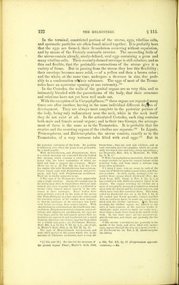
![the genital organs of the first is yet imperfectly known; while that of those of the second is well understood. The female apparatus of the Tremato- des consists of a germ-forming organ (ovary), with its excretory duct; then, two others for forming the vitellus, which have also excretory ducts; and then a simple uterus with its vagina. The male apparatus con- sists of testicles with their excretory canals, an internal seminal vesicle, a cirrhus-sac, an external seminal vesicle, and a penis.® The ovary consists of a round or pyriform ® reservoir, situated, usually, upon the median line of the body,® from which it is distinguished by its pale color and transparency. It is filled with simple round cells — the egg-germs. The nucleus of these cells is the germinative vesicle, and the nucleolus, the germinative dot.® The short and small excretory duct of the ovary opens at the commence- ment of the uterus. The organs which secrete the vitellus are two in num- ber, of variable length, and situated upon each side of the body near the dorsal surface; they occupy either the cervical, the central, or the posterior portion of the animal, and sometimes extend over them all. They are nearly always composed of ramified caeca filled with white, granular, vitelline corpuscles. By reflected light these caeca appear through the skin as a white, ramified, botryoidal mass,® and from each of them, pass off inwardly, numerous excretory ducts, which reunite opposite the ovary into two common canals. These last approach each other transversely, and form a single canal upon the median line, which, after a short course, opens at the bottom of the uterus by an orifice which is common to it and the ovary.® Pentastomum taenioides, organs which are re- garded by Diesing as caeca for secreting the en- velope of the eggs. Since all the parts of the genital organs of Pen- tastomum have not been examined with this same precision, I can give no opinion as to their use.* 2 See Siebold, in Wiesmann's Arch. 1836, I. p. 217, Taf. VI., and in Muller's Arch. 1836, p. 232, Taf. X. fig. 1. 3 The ovary here is always smaller than the testicle, and sometimes as to form very closely resembles it, as in Distomum globiporum, and longicolle, mihi (from the urinary bladder of Cottus gobio); consequently it may easily be taken for a third testicle. 4 With Monostomum, it lies wholly at the pos- terior extremity. 5 In Polystomum, Octobothrium and Diplo- zoon, the germs are so large that they may easily be taken for perfect eggs. There is here, moreover, between the cell-wall and the nucleus (the germinative vesicle), quite a thick layer of albuminous substance, somewhat representing a vitellus. But in the other Trema- todes it is so thin as scarcely to be perceived. 0 With the following Trematodes there is a wide deviation from this usual arrangement. In Dis- tomum longicolle the organs producing the vitellus are two simple round caeca located behind the ventral sucker ; in Distomum cygnoides, they are two very small deeply-fissured bodies j and in Distomum gibbosum, there is one only, which is star-shaped and located nt the middle of the body. 7 These organs, until now regarded as ovaries, secrete only vitelline cells. With most Trematodes their nuclei are clear, and have been taken for eggs. In eggs recently formed, one can always distinguish these cells from the germs. In passing the excretory canals they are compressed and elongated, but never run into each other. When these canals are crowded, they have the aspect of white cords, which have often been taken for nerves. But when they are empty, they, as well as the vitellus-segreting organs, are almost invis- ible.! * [ § 115, note 1.] See upon this subject Van Beneden (Ann. d. Sc. Nat. XI. 1849, p. 326), who has described in detail the sexual organs of Lin- guatula Diesingii, and has shown the sexes to be separate. See also my note under § 99. — Ed. t[§ 115, note 7.] To say that certain organs secrete vitelline cells, is a little obscure, and no doubt Siebold intended to convey the meaning that they secreted the plastic material out of which these cells are formed. I make _ this perhaps seemingly unnecessary reference to the matter, since it concerns the subject of the development of the ovum. In the Ascaris, where the origin and development of the ovum can be satisfactorily studied, you first notice the germs as nucleolated cells, of which the nucleus is the future germina- tive vesicle and the nucleolus the germinative dot. These cells jncrease in size, and as they move along there appear in the liquid which lies between the nucleus and the cell-wall minute granules which ultimately become cells ; in this way the vitellus is formed, the formation being endogenous and not exogenous. These special organs or tubes therefore are vitellus-forming organs, in vir- tue of their secreting the formative material out of which the vitellus is formed within the original, nucleolated germ-cell. — Ed.](https://iiif.wellcomecollection.org/image/b2491874x_0124.jp2/full/800%2C/0/default.jpg)