A microscopic examination of the ova of Penthaleus capidarius / by T. Spencer Cobbold, Esq., M.D. (Senior President of the Royal Medical Society of Edinburgh).
- Thomas Spencer Cobbold
- Date:
- 1852
Licence: Public Domain Mark
Credit: A microscopic examination of the ova of Penthaleus capidarius / by T. Spencer Cobbold, Esq., M.D. (Senior President of the Royal Medical Society of Edinburgh). Source: Wellcome Collection.
Provider: This material has been provided by The University of Glasgow Library. The original may be consulted at The University of Glasgow Library.
4/8 (page 2)
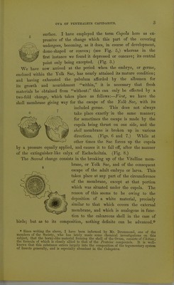
![can at once detect their form and recognise their nature. Their diminu- tive size is a character common to most species of the same family, (Acaridce.) To obtain, however, a more satisfactory knowledge of their morphology and construction, it is necessary to subject them to much higher magnifying powers, and the drawings now before the Society, represent them at different stages of development, magnified two hundred and fifty diameters linear. The chief points of interest, to which we may direct our attention, bear reference to their external configuration; the changes in form during development; and the manner in which the mature embryo or larva makes its escape from the outer covering or shell. The general form then may be at once recognised as that of a sphere flattened and depressed from above. (See Figs. 2 and 3.) This button-like character is by no means an unfrequent appearance in the eggs of insects generally; and though I have not seen any precisely similar illustrations to those before us in the works of recent entomological writ- ers; there is, nevertheless, figured in the ereat work of Swammerdam, Plate xxxiii, Fig. 1, a very similar form of an egg, belonging to one of the Noctme, or Post Meridian Moths; it is devoid! however, of certain markings, which, in the specimens before us, have a very chaste appearance, and which represent a series of alternate elevations and depressions, twenty-five in number, running from a central elevated point, to the outer border of the button-like surface. When viewed laterally, and especially if the specimen be immersed in some transparent medium, not only are these morphological features readily confirmed, but we observe, through the outer covering or 7 shell-membrane, a small round opaque body, situated im- 1 mediately under the central prominence, as seen in Fig. 4- This there can be little doubt, corresponds to the Yolk, or Vitellus, in the Ova of birds and other animals, being enclosed in a special and distinct envelope of its own, con- stituting the Yolk Sac, or Vitelline membrane. Keeping our attention fixed from time to time on the same Ova we observe that this opaque body or Yolk increases a h expose of the surrounding medium or albumen, which is situated W n it and the shell membrane, the alterations in form of which ]2T tructure are almost entirely confined to the cupola, or cup-shaped](https://iiif.wellcomecollection.org/image/b21464935_0004.jp2/full/800%2C/0/default.jpg)