Licence: Public Domain Mark
Credit: A system of medicine / by many writers. Source: Wellcome Collection.
Provider: This material has been provided by UCL Library Services. The original may be consulted at UCL (University College London)
69/944 page 45
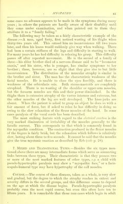
![is primarily interstitial, and consists of an overgrowth of fat and nucleated fibrous tissue between the muscle-fibres, and secondarily in the muscle- fibres, which become narrow and irregular in shape. The transverse striation is at first preserved, but it becomes more faint, and finally disappears, and the muscle undergoes a granular degeneration. Earely the muscle-fibres shew fatty degeneration, longitudinal striation, fissuring, vitreous (waxy) degeneration, or vacuolation (Gowers). According to Duchenne, the sheaths of the sarcolemma appear to contain fat-cells which are really derived from the surrounding connective tissue, and which otherwise differ from the fatty graiuilar condition characteristic of Fifi. 10.—Transverse section nf a i)Ortion of ]ts<Mi<lo-liyi)ertroitliic iniisfl«! shewing; tin- typical changes,, viz. atiopliy of miiscle-libres, hypertrophy of soiiu- lihrrs, iiieicase of intfrniiiscular connective tissue, increase of nuch'i, ami excess of atliposc tissue within tin- ninsclr. Majjiiilied 30 diameters ; the lar^,'e fibres measure Q-l nun. in diametfr. The normal nuiscle-libres of an adult measure •Otl mm., and those of a hoy »'i;;ht years old nun. fatty degeneration of muscle ; and the interstitial connective tissue is not produced by fibroid changes of the muscle. Ill a late case of pseudo-hypertrophic myopathy Ballet and Laignel- Lavastine found the characteristic changes in tlie muscles, and that the })eripheral nerves did not shew any indication of a neuritic process in the cervical enlargement. In the spinal cord there was almost com2:)Iete absence of the antei ior cornual cells ; such cells as persisted were not deformed but simply atrophic, there being no sign of chromatolysis, pigmentation, or neuroglial proliferation. In the motor cortex the Betz- cells were usually glol)ular and deformed. The atrophy of the cells was regarded as secondai v to the muscular chanii;e. The case on whicli Landouzy and Dejerine based their original descrip- tion of the facio-scapulo-humeral type in 1874 died in 1902 at the age of forty-five years ; the pathological examination shewed the most advanced atrophy of the muscles, some, such as the biceps, being very small and of](https://iiif.wellcomecollection.org/image/b21274083_0069.jp2/full/800%2C/0/default.jpg)


 No text description is available for this image
 No text description is available for this image
 No text description is available for this image