Licence: In copyright
Credit: The muscles of the eye / by Lucien Howe. Source: Wellcome Collection.
Provider: This material has been provided by UCL Library Services. The original may be consulted at UCL (University College London)
56/504 page 36
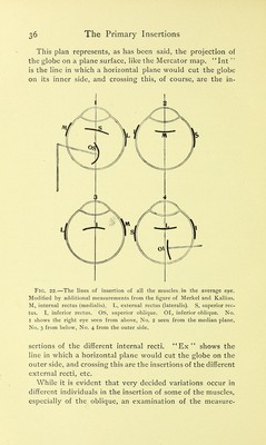
![This plan represents, as has been said, the projection of the globe on a plane surface, like the Mercator map. Int is the line in which a horizontal plane would cut the globe on its inner side, and crossing this, of course, are the in- Fig. 22.—The lines of insertion of all the muscles in the average eye. Modified by additional measurements from the figure of Merkel and Kallius. M, internal rectus (medialis). L, external rectus (lateralis). S, superior rec- tus. I, inferior rectus. OS, superior oblique. 01, inferior oblique. No. I shows the right eye seen from above, No. 2 seen from the median plane, No. 3 from below, No. 4 from the outer side. sertions of the different internal recti. Ex shows the line in which a horizontal plane would cut the globe on the outer side, and crossing this are the insertions of the different external recti, etc. While it is evident that very decided variations occur in different individuals in the insertion of some of the muscles, especially of the oblique, an examination of the measure- ] ?](https://iiif.wellcomecollection.org/image/b21287004_0056.jp2/full/800%2C/0/default.jpg)


 No text description is available for this image
 No text description is available for this image
 No text description is available for this image