Collected papers on trypanosomiasis / by Sir David Bruce and others.
- David Bruce
- Date:
- [1909-1911]
Licence: Public Domain Mark
Credit: Collected papers on trypanosomiasis / by Sir David Bruce and others. Source: Wellcome Collection.
45/268 (page 390)
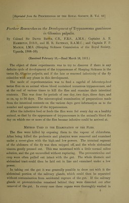
![[Reprinted from the Proceedings of the Royal Society, B. Yol. 83] Further Researches on the Development of Trypanosoma gambiense in Glossina palpalis. By Colonel Sir David Bruce, C.B., F.R.S., A.M.S.; Captains A. E. Hamerton, D.S.O., and H. R. Bateman, R.A.M.C.; and Captain F. P. Mackie, I.M.S. (Sleeping Sickness Commission of the Royal Society, Uganda, 1908-10). (Received February 15,—Read March 16, 1911.) The object of these experiments was to try to discover if there is any definite cycle of development of the trypanosome of Sleeping Sickness in the tsetse fly, Glossina 'palpalis, and if the late or renewed infectivity of the fly coincides with any phase in this development. The mode of experimentation was to feed a cageful of laboratory-bred tsetse flies on an animal whose blood contained numerous trypanosomes, and at the end of various times to kill the flies and examine their intestinal contents. This was done for periods of one day, two days, three days, and so on, up to 56 days. The microscopical examination of preparations made from the intestinal contents on the various days gave information as to the number and appearance of the trypanosomes. After the infective feed or feeds the flies were fed every day on a healthy animal, so that by the appearance of trypanosomes in the animal’s blood the day on which one or more of the flies became infective could be arrived at. Method Used in the Examination of the Flies. The flies were killed by exposing them to the vapour of chloroform. After being killed the proboscis and pharynx were removed and examined under a cover-glass with the high, and low powers. The terminal segment of the abdomen of the fly was then snipped off, and the whole abdominal viscera gently pressed out. This was moistened with a little normal saline solution, and the gut unravelled without rupturing. The proventriculus and crop were often pulled out intact with the gut. The whole thoracic and abdominal tract could then be laid out in line and examined under a low power. In taking out the gut it was generally possible to draw out with it the abdominal portion of the salivary glands, which could then be separated without contamination from accidental rupture of the gut. If the salivary glands or proventriculus remained behind they were dissected out after removal of the gut. In every case these organs were thoroughly washed in b](https://iiif.wellcomecollection.org/image/b31360841_0045.jp2/full/800%2C/0/default.jpg)