Obstetrics : the theory and practice ; including the diseases of pregnancy and parturition, obstetrical operations, etc. / by P. Cazeaux ; remodelled and rearranged, with additions and revisions, by S. Tarnier.
- Pierre Cazeaux
- Date:
- 1885
Licence: Public Domain Mark
Credit: Obstetrics : the theory and practice ; including the diseases of pregnancy and parturition, obstetrical operations, etc. / by P. Cazeaux ; remodelled and rearranged, with additions and revisions, by S. Tarnier. Source: Wellcome Collection.
Provider: This material has been provided by The University of Leeds Library. The original may be consulted at The University of Leeds Library.
39/1140 (page 35)
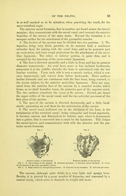
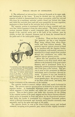
![is so well marked as to be mistaken, when practising the touch, for the Bacro-vertebral angle. The anterior sacral foramina, four in number, are found nearer the lateral margins; they communicate with the sacral canal, and transmit the anterior branches of the nerves of the same name. Beyond the foramina is an unequal surface for the attachment of the pyramidal muscles. 3. The borders of the sacrum may be divided into two portions. 1. The superior, being very thick, presents, on its anterior half, a semilunar articular facet for joining with the coxal bone, and on its posterior part an excavation, and some rough projections for the attachment of the sacro- iliac ligaments. The other, or inferior portion, is quite thin, and is occupied by the insertion of the sacro-sciatic ligaments. 4. The base is directed upwardly and a little in front, and has its greatest diameter transversely. An oval facet, more or less inclined backwards, surmounts it at the middle, whereby the bone is articulated with the last lumbar vertebra. Upon each side is seen a smooth surface, which is con- cave transversely, and convex from before backwards. These surfaces incline forwards and are continuous with the iliac fossae, being covered, in the recent subject, by the anterior sacro-iliac ligaments. They are sepa- rated from the anterior face of the sacrum by a rounded border, which forms, as we shall hereafter learn, the posterior part of the superior strait. The two surfaces constitute the ivings of the sacrum. Behind, are found the upper orifice of the sacral canal, and the two articular processes of the first piece of the sacrum. 5. The apex of the sacrum is directed downwards, and a little back- wards; presenting an oval facet for the articulation of the coccyx. 6. The sacral canal, hollowed out in the thickness of the bone, is the termination of the vertebral canal; being triangular and broad superiorly, it becomes narrow and flattened at its inferior part, where it degenerates into a gutter, that is converted into a canal by the ligaments. This lodges the sacral nerves, and communicates both with the anterior and the pos- terior sacral foramina. FiQ. 1. Fig. 2. B B D ]J Anterior surface of the sacrum. Posterior surface of the sacrum. Fio. 1. A. Ala or wings of the sacrum. B. Articular processes. C. Anterior sacral foramina. E. Points «if attachment of the right pyramidal muscle. Fio 2. A. Ridge formed by the spinous processes. B. Posterior sacral foramina. D. Articular processes. The sacrum, although quite thick, is a very light and spongy bone. Besides, it is pierced by a great number of foramina, and traversed by a central cavity, which serve to diminish its weight still more.](https://iiif.wellcomecollection.org/image/b21515013_0041.jp2/full/800%2C/0/default.jpg)