Obstetrics : the theory and practice ; including the diseases of pregnancy and parturition, obstetrical operations, etc. / by P. Cazeaux ; remodelled and rearranged, with additions and revisions, by S. Tarnier.
- Pierre Cazeaux
- Date:
- 1885
Licence: Public Domain Mark
Credit: Obstetrics : the theory and practice ; including the diseases of pregnancy and parturition, obstetrical operations, etc. / by P. Cazeaux ; remodelled and rearranged, with additions and revisions, by S. Tarnier. Source: Wellcome Collection.
Provider: This material has been provided by The University of Leeds Library. The original may be consulted at The University of Leeds Library.
74/1140 (page 70)
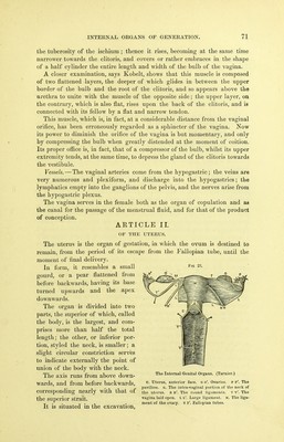
![The inferior extremity, or vulvar orifice, presents, in front, a transvereo rugous prominence, that seems to diminish the entrance. Structure of the Vagina. — [The walls of the vagina average in thickness from one-eighth to three-sixteenths of an inch. It is composed of thi-ee layers: one external or cellulo-fibrous; a middle or muscular one ; and the internal or mucous. The external layer is composed of fibres of both elastic and connective tissue; it blends externally with the organs surrounding the vagina, and internally with the middle layer. The middle layer is composed of muscular fibres which are inserted in front upon the branches of the ischium and pubis, and are continued upward to become blended with the middle layer of fibres of the uterus. Some again disappear upon the utero sacral ligaments, whilst others cross each other in all directions, leaving interspaces occupied by projecting veins. The internal or mucous layer is of a pale-red color, which becomes violet during menstruation and especially during pregnancy. Its external surface is con- founded with the' preceding layer, whilst its internal is covered with tessellated epithelium and abounds in folds analogous to papillae. For a long time this mem- brane was supposed to be rich in mucous follicles, but anatomists now agree in tho opinion that the vagina is destitute of mucous glands. In great part, the walls of the vagina are composed of a tissue possessing all the characters of spongy erectile tissue; that such is the case has been proved beyond cavil by the researches of M. Kobelt and Ch. Eouget.] According to Kobelt, this erectile tissue is composed of several superposed layers of venous network which proceed from the bulb, the finest ramifica- tions extending into the mucous membrane. This true spongy body extends continuously through the entire limits of the vestibule and of the vagina, and seems connected with the veins of the parenchyma of the uterus. The great vascularity of the walls of the vagina explains, to a certain extent, the dangers consequent upon their rupture. Surrounding the lower extremity of the vagina are a few muscular fibres, that constitute what is erroneously called (see below) the constrictor vaginae muscle. In some females, this is quite strong and well developed. Finally, under the name of bulb of the vagina, a swelling or cavernous body is described, that separates the orifice of this canal from the roots of the clitoris: moderately thick in the centre, where it is placed between the meatus urinarius and the junction of the crurse clitoridis, it gradually swells out, as it recedes from this point, and terminates below in an enlarged extremity on the sides of the vagina, being deficient, however, on the posterior wall of this canal. The length of the bulb, when injected, is about one inch and three-eighths of an inch; its greatest width, from one-half to three-quarters of an inch, and its thickness from about three-eighths of an inch to one-quarter of an inch. (Kobelt.) The bulb of the vagina is com- posed of an erectile tissue analogous to that of the bulb of the urethra in the male, and communicates freely, as shown by M. Deville, with the cavernous tissue of the clitoris, by means of several veins of considerable size. The bulb of the vagina is surrounded, as it were, by a layer of muscular fibres {constrictor cunni), in regard to the arrangement of which authors difier. According to Kobelt, there are two constrictor muscles. It takes its origin by a large and flattened base from the aponeurosis of the perineum, alm()St directly at the middle of the space which separates the anus fronj](https://iiif.wellcomecollection.org/image/b21515013_0076.jp2/full/800%2C/0/default.jpg)