Obstetrics : the theory and practice ; including the diseases of pregnancy and parturition, obstetrical operations, etc. / by P. Cazeaux ; remodelled and rearranged, with additions and revisions, by S. Tarnier.
- Pierre Cazeaux
- Date:
- 1885
Licence: Public Domain Mark
Credit: Obstetrics : the theory and practice ; including the diseases of pregnancy and parturition, obstetrical operations, etc. / by P. Cazeaux ; remodelled and rearranged, with additions and revisions, by S. Tarnier. Source: Wellcome Collection.
Provider: This material has been provided by The University of Leeds Library. The original may be consulted at The University of Leeds Library.
85/1140 (page 81)
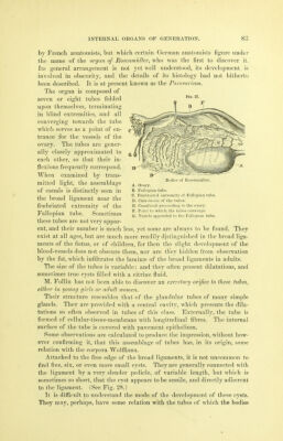
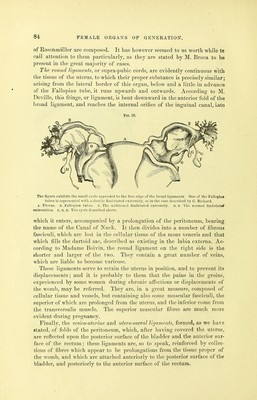
![2. The glands of the neck (glandS; or ovula of Naboth) are found in al] the interval between the line separating the cavity of the neck from that of the body, and the neighborhood of the borders of the os tincse. Their orifices are readily seen upon, and especially between, the folds of the arbor vitse. These glands have the form of a minute cylinder, terminating in a rounded cul-de-sac, which is inflated into the form of a lentil or vial, and inclosed in the tissue of the mucous membrane, even descending a little between the fibres of the muscular structure. The excretory orifice is always smaller than the glandular tube. Pres- sure causes the escape from it of a transparent, viscid, tenacious, and com- pletely homogeneous fluid. We shall treat hereafter of the modifications which these glands undergo during gestation. [The epithelium of the uterine mucous membrane is cylindrie, with vibratile cilia moTing from without inward. It is therefore impossible that the ciliary motion should carry the spermatic fluid toward the openings of the tubes, as has been erroneously supposed. The entire cavity of the body and of the neck, to a point near the external ori- fice of the latter, is covered with vibratile epithelium. Below this point the mucous membrane of the neck is furnished with the pavimentous variety. D. Vessels.—The arteries of the uterus proceed from the hypogastric and ovarian arteries. Both present many flexuosities in their course through the tissue of the organ, and are remarkable for their corkscrew form, recalling the arrangement of the helicine arteries. The neck is less vascular than the body. The veins are highly developed, anastomosing freely, and forming cavities, as it were, in the muscular tissue. They are called uterine sinuses, and communicate largely with the venous plexuses within the folds of the broad ligaments. From the latter proceed the uterine and ovarian veins which empty into the correspond- ing trunks. From the arrangement of the uterine arteries and veins, surrounded as they are everyvi'here by muscular partitions, it results, that the uterus is a true erectile organ, as has been placed beyond doubt by an excellent memoir published by Professor Rouget. This skilful anatomist has, in fact, shown that by injecting the veins of the uterus the organ is put in a state of true erection, whereby it rises, swells, and moves up toward the abdomen. Under these circumstances its volume is greater by one-half than in the empty condition, and the walls of the cavity separate from each other. These phenomena doubtless take place during coition, and probably facilitate the ascent of the spermatic fluid. The lymphatic vessels are very abundant, and pass into the pelvic and lumbar ganglia. E. Nerves.—The nerves are derived from the great sympathetic, some of them pro- i!eeding from the renal and others from the hypogastric plexuses; to the latter are united some fibres from the sacral plexus.] It is an important practical remark of M. Jobert, that the entire intra- vaginal portion of the neck is destitute of a supply of nervous fibres, whilst the portion above the insertion of the vagina receives a great numl)er of them, which form species of plexuses, furnishing ascending or uterine- 6](https://iiif.wellcomecollection.org/image/b21515013_0087.jp2/full/800%2C/0/default.jpg)