Obstetrics : the theory and practice ; including the diseases of pregnancy and parturition, obstetrical operations, etc. / by P. Cazeaux ; remodelled and rearranged, with additions and revisions, by S. Tarnier.
- Pierre Cazeaux
- Date:
- 1885
Licence: Public Domain Mark
Credit: Obstetrics : the theory and practice ; including the diseases of pregnancy and parturition, obstetrical operations, etc. / by P. Cazeaux ; remodelled and rearranged, with additions and revisions, by S. Tarnier. Source: Wellcome Collection.
Provider: This material has been provided by The University of Leeds Library. The original may be consulted at The University of Leeds Library.
96/1140 (page 92)
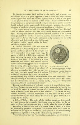
![a single mass. I have seen the yolk divided in two, and, on one occasion, into five parts of different volume. The vitellus usually fills the interior of the zone completely, and has the same foi*m, but sometimes the vitelline sphere is smaller than that destined to receive it. Some authors likewise believe that a very delicate membrane exists, which incloses and unites the yolk in a single mass: but Messrs. Coste and BischofF agree in rejecting the existence of this, and contend that the granulations of the vitellus are placed in juxtaposition with the transparent zone, whioh forms its sole and only envelope. c. Germdnal Vesicle. — In the midst of the vitellus, in very young girls, or on one of the neighboring points of the peripheral envelope in the matured ovules, a small, perfectly transparent, and colorless vesicle is seen like a clear spot, surrounded by a mass of a deeper yellow. Purkinje had described it in the eggs of birds, and gave his own name to it; but M. Coste is entitled •to the honpr of Jiaving first demonstrated its existence in the ovum of mammiferie, and of*thus having established the perfect identity between the : latter and the eggs of birds. This is the vesicle of Purkinje, or the germinal vesicle. It is slightly oval, and consists of a very delicate, transparent, and colorless membrane, which incloses a liquid that is frequently as limpid and transparent as itself, though it sometimes contains a few granules. Notwith- standing its extreme tenuity, this vesicle still offers a certain consistence, since it has been seen intact, after leaving the ovule, and being completely separated from the granular liquid in which it was placed. It is always very small, and scarcely measures the sixtieth of a line in diameter. D. The Germinal Spot. — If the germinal vesicle be attentively observed, an obscure rounded spot will be seen on some part of its periphery; this was first discovered by Wagner, who gave it the name of the germinal spot. It seems to be formed by the aggregation of fine small granules, or little globules, the obscure hue of which is brought out by the clear contents of the vesicle. Wagner has sometimes met with two, or even more, germinal spots in the mammiferse. Before fecundation, therefore, the ovule is composed: 1st, of an exterior envelope, the vitelline membrane, or transparent zone; 2d, of a vitellus, or yolk, contained in this vesicle; 3d, of a little vesicle inclosed in the first and swimming in the vitelline fluid — the germinal vesicle; 4th, and lastly, of the germinal spot. EXPLANATION OF PLATE 1. MEDIAN PERPENDICULAR SECTION OF PELVIS FROM FRONT TO BACK, SHOWING BOTH PELVIC SPACES. \_Taken from Savage on the Female Pelvic Organs.] A. Anus, marking the columns of Morgagni. r. Rectum, projections in the cavity, the valves (?) of Houston. These folds include all the coats of the rectum, and are readily effaceable by slight distension. Note minute circular markings at the anal end, indicating transverse sections of the inferior circular fibres of the rectum (interniil sphincter), and lines near the coccyx indicating the posterior half of external sphinc- ter, the coccygeal attachment of the pubo-coccygeal muscle, and the recto-coccygeus](https://iiif.wellcomecollection.org/image/b21515013_0098.jp2/full/800%2C/0/default.jpg)