Tobacco amblyopia in a woman, with anomalous scotomas / by G. E. de Schweinitz.
- De Schweinitz, G. E. (George Edmund), 1858-1938.
- Date:
- [1897]
Licence: Public Domain Mark
Credit: Tobacco amblyopia in a woman, with anomalous scotomas / by G. E. de Schweinitz. Source: Wellcome Collection.
Provider: This material has been provided by UCL Library Services. The original may be consulted at UCL (University College London)
4/10 (page 2)
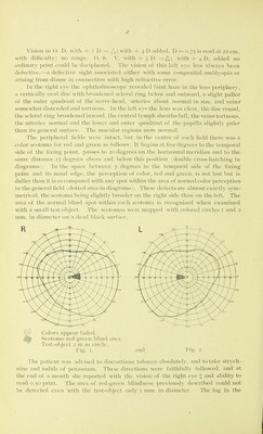
![\'isi(^n in (). D. with i D = /o; with -h 4 D added, T) =0.75 is read at 22 cm. with difficulty; no ran^'e. (_). S. \'. with ^ 3 D. =,,';,; with — 4 1). added no ordinary jndnt coidd l)e deciphered. Tlie vision of this left e\-e has alwa\-s been defective.—a defectixe si^lit associated either with some congenital amljh'opia or arising from disuse in connection with high refractive error. In the right eye the ophthalniosco])e revealed faint haze in the lens peripherv, a vertically oval disc with broadened scleral ring below and outward, a slight pallor of the outer quadrant of the nerve-head, arteries about normal in size, and veins somewhat distended and tortuous. In the left eye the lens was clear, the disc round, the scleral ring broadened inward, the central lymph .sheaths full, the veins tortuous, the arteries normal and the lower and outer quadrant of the papilla slighth- paler than its general surface. The macular regions were ncjrmal. The peripheral fields w-ere intact, but in the centre of each field there was a color scotoma for red and green as follows: It begins at five degrees to the temporal side of the fixing point, passes to 20 degrees on the horizontal meridian and to the same distance 15 degrees, above and below this position 1 double cross-hatching in diagrams). In the space between 5 degrees to the temporal side of the .fixing point and its nasal edge, the perception of color, red and green, is not lo.st but is diiller than it is as compared with any spot within the area of normal color perception in the general field (dotted area in diagrams). These defects are alnio.st exactlv sym- metrical, the scotoma being slightly broader on the right side than on the left. The area of the normal blind .spot within each scotoma is recognized when examined with a small test-object. The scotomas were mapped with colored circles i and 2 mm. in diameter on a dead black surface. Colors ap])ear faded. Scotoma re'd-green blind area. Test-object 2 ni ni circle. I'ig. I. I-i< The ])atient was adN ised to discnUnue tobacco al)solutel\-. and to take strych- nine and iodide of ])otassiuni. 'Hiese directions were faithfuU}' followed, and at the end of a month she re])orted with the vision of the right eye I; and ability to read 0.50 print. The arc-a of red-green blindness previously descri])ed could not be detected even \\\\\\ tlie test-ol»jecL only 1 nun. in diameter. The fog in the](https://iiif.wellcomecollection.org/image/b21648554_0004.jp2/full/800%2C/0/default.jpg)