Congenital dermal sinuses : a source of spinal memnigeal infection and subdural abcesses / by A. Earl Walker and Paul C. Bucy.
- Arthur Earl Walker
- Date:
- [1934?]
Licence: In copyright
Credit: Congenital dermal sinuses : a source of spinal memnigeal infection and subdural abcesses / by A. Earl Walker and Paul C. Bucy. Source: Wellcome Collection.
22/26 (page 418)
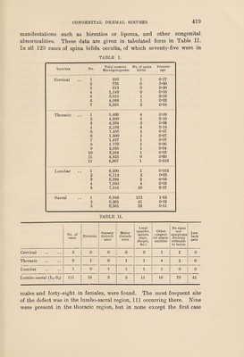
![dermal sinus which, unlike the condition found in the other cases, passed through the opening normally present between the laminae of the 4th and 5th lumbar vertebrae. Thus, although it can be stated that this peculiar malformation is usually associated with a spina; bifida it is not necessarily so. Although spina bifida occulta is generally considered as of not infrequent occurrence, there are very few statistics on which to base this conclusion. Mayer [4] states that a complete splitting of the sacrum occurred in 3 per cent, of cases, and that in children over 10 years of age the first sacral lamina was not fused in 24 per cent, of cases in the general population. These figures indicate a much higher incidence of spina bifida than that reported by Sutherland [9] at the Mayo Clinic. In approximately 12,000 roentgenograms of the spine he found 621 cases of spina bifida, an incidence of slightly more than 5 per cent. The condition was twice as common in males as in females. It involved the 1st and 2nd sacral vertebrae in 70 per cent, of cases, the 5th lumbar in 24'5 per cent. No mention is made of the incidence of spina bifida occulta in the thoracic region, but four cases are presented with the defect in that region. In 1920 Theodora Wheeler reviewed the literature to that time, and in a group of 1,000 consecutive X-ray plates of the sacrum found the incidence of spina bifida occulta of the first sacral vertebra to be 13'1 per cent., and the laminae of the entire sacrum were unfused in 2'89 per cent, of cases. The 5th lumbar was found unfused in 23 cases of the 1,000, and the atlas, the only other vertebra in that series with spina bifida, in 1'47 per cent. Table I shows the incidence of spina bifida occulta in a series of roentgenograms of ‘the spine taken at the University of Chicago Clinics in the past six years. These include films taken to show trachea, cervical spine, colon, chest, thoracic spine, lumbar spine, urinary tract, pelvis, sacro-iliac joints, and lumbo-sacral spine; some of which only incidentally showed the vertebrae. The total number of patients of whom roentgenograms of these parts were taken was noted, and then twenty representative roentgenograms of each studied to determine the mean number of vertebrae included in any roentgenogram of any of the above structures. From these data it was a simple mathematical problem to calculate the number of times each vertebra had been shown in the total number of roentgenograms. All cases of spina bifida occulta were then collected and reviewed to determine the location of the anomaly, its association with enuresis, motor or sensory defects, local](https://iiif.wellcomecollection.org/image/b30629871_0022.jp2/full/800%2C/0/default.jpg)