Licence: Public Domain Mark
Credit: Hand-book of physiology / by W. Morrant Baker and Vincent Dormer Harris. Source: Wellcome Collection.
Provider: This material has been provided by the Royal College of Physicians of Edinburgh. The original may be consulted at the Royal College of Physicians of Edinburgh.
102/930 (page 74)
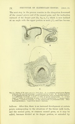
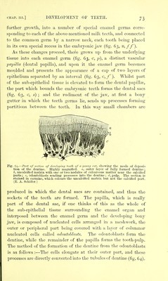
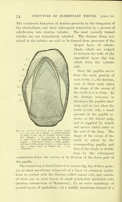
![The contimied formation of dentine proceeds by the elongation of the odontoblasts, and their snbsequent convei'sion hj a process of calcification into dentine tnbnles. The most recently formed tubules are not immediately calcified. The dentine fibres con- tained in the tidiides are said to be formed from processes of the deeper hiyer of odonto- blasts, which are wedged in between the cells of the superficial layer (fig. 64) which form the tubules only. Since the papillaj are to form the main portion of each tooth, i.e., the dentine, each of them early takes the shape of the crown of the tooth it is to form. As the dentine increases in thickness, the jjapillte dimi- nish, and at last when the tooth is cut, only a small amount of the papilla re- mains as the dental pulp, and is supplied by vessels and nerves which enter at the end of the fang. The shape of the crown of the tooth is taken by the corresponding papilla, and that of the single or double fang by the subsequent constriction below the crown, or by division of the lower part of the papilla. The enamel ca]j is found later on to consist (fig. 65) of three parts: (a) an inner membrane, composed of a layer of columnar epithe- lium in contact with the dentine, called enamel celh, and outside of these one or more layers of small polyhedral nucleated cells (stratum intermedmm of Hannover); (h) an outer membrane of several layers of epithelium ; (c) a middle membrane formed of a Fig. 65.—Vertical trinisverse section of the dentnl sac, 2'iilp, ice, of a kitten, n, dental papilla or pulp ; b, the cap of dentine formed upon the summit; c, its covering of enamel; d, inner layer of epithelium of the enamel organ ; e, ;,''elatinou.s tissue; /, outer epithe- lial layer of the enamel organ; //, inner layer, and A, outer layer of dental sac. X 14. (Thiersch.)](https://iiif.wellcomecollection.org/image/b21906300_0104.jp2/full/800%2C/0/default.jpg)