Licence: Public Domain Mark
Credit: Hand-book of physiology / by W. Morrant Baker and Vincent Dormer Harris. Source: Wellcome Collection.
Provider: This material has been provided by the Royal College of Physicians of Edinburgh. The original may be consulted at the Royal College of Physicians of Edinburgh.
57/930 (page 29)
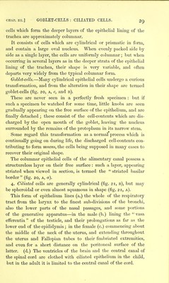
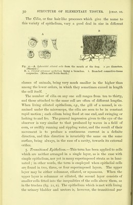
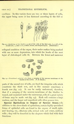
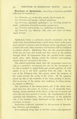
![CHAP. III.] GOBLET-CELLS : CILIATED CELLS. cells which form the deeper layers of the epithelial lining of the trachea are approximately columnar. It consists of cells which are cylindrical or prismatic in form, and contain a large oval nucleus. When evenly packed side by side as a single layer, the cells are uniformly columnar; but when occurring in several layers as in the deeper strata of the epithelial lining of the trachea, their shape is very variable, and often departs very widely from the typical columnar form. Goblet-cells.—Many cylindrical epithelial cells undergo a curious transformation, and from the alteration in their shape arc termed goblet-cells (fig. 20, a, c, and b). These are never seen in a perfectly fresh specimen : but if such a specimen be watched for some time, little knobs are seen gradually appearing on the free surface of the epithelium, and are finally detached ; these consist of the cell-contents which are dis- charged by the open mouth of the goblet, leaving the nucleus surrounded by the remains of the protoplasm in its naiTow stem. Some regard this transformation as a normal process which is continually going on during life, the discharged cell-contents con- tributing to form mucus, the cells being supposed in many cases to recover their original shape. The columnar epithelial cells of the alimentary canal possess a stnxctureless layer on their free surface : such a layer, appearing striated when viewed in section, is termed the striated basilar border (fig, 20, a, a). 4. Ciliated cells are generally cylindrical (fig. 21, b), but may be spheroidal or even almost squamous in shape (fig. 21, a). This form of epithelium lines (a.) the whole of the respiratory tract from the larynx to the finest sub-divisions of the bronchi, also the lower parts of the nasal passages, and some portions of the generative apparatus—in the male (b.) lining the vasa efferentia of the testicle, and their prolongations as far as the lower end of the epididymis; in the female (c.) commencing about the middle of the neck of the uterus, and extending throughout the uterus and Fallopian tubes to their fimbriated extremities, and even for a short distance on the peritoneal surface of the latter, (d.) The ventricles of the brain and the central canal of the spinal cord are clothed with ciliated epithelium in the child, but in the adult it is limited to the central canal of the cord.](https://iiif.wellcomecollection.org/image/b21906300_0057.jp2/full/800%2C/0/default.jpg)