Licence: Public Domain Mark
Credit: Hand-book of physiology / by W. Morrant Baker and Vincent Dormer Harris. Source: Wellcome Collection.
Provider: This material has been provided by the Royal College of Physicians of Edinburgh. The original may be consulted at the Royal College of Physicians of Edinburgh.
72/930 (page 44)
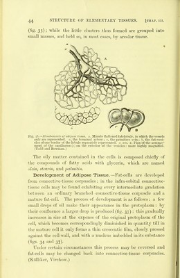
![(fig. 3S); while the little clusters thus formed are grouped into small masses, and held so, in most cases, by areolar tissue. Fig. 36.—Blood-vessds of adipose tissue. A. Minute flattened fat-lobule, in which the vessels only are represented, n, the terminal artery ; y, the primitive vein; b, the fat-vesi- cles of one border of the lobule separately represented, x loo. b. Plan of the arrange- ment of the capillaries (c) on the exterior of the vesicles: more highly magnifled. (Todd and Bowman.) The oily matter contained in the cells is composed chiefly of the compounds of fatty acids with glycerin, which are named olein, stearin, and palmitiii. Development of Adipose Tissue.—Fat-cells are developed from connective-tissue corpuscles : in the infra-orbital connective- tissue cells may be found exhibiting every intermediate gradation between an ordinary branched connective-tissue corpuscle and a mature fat-cell. The process of development is as follows : a few small drops of oil make their appearance in the protoplasm : by their confluence a larger drop is produced (fig. 37) : this gradually increases in size at the expense of the original protoplasm of the cell, which becomes correspondingly dimhiished in quantity till in the mature cell it only forms a thin crescentic film, closely pressed against the cell-wall, and with a nuclens imbedded in its substance (figs. 34 and 37). Under certain circumstances this process may be reversed and fat-cells may ])e changed back into connective-tissue corpuscles. (Kolliker, Virchow.)](https://iiif.wellcomecollection.org/image/b21906300_0072.jp2/full/800%2C/0/default.jpg)