A treatise on mental diseases : based upon the lecture course at the Johns Hopkins University, 1899, and designed for the use of practitioners and students of medicine / by Henry J. Berkley.
- Henry Johns Berkley
- Date:
- 1901
Licence: In copyright
Credit: A treatise on mental diseases : based upon the lecture course at the Johns Hopkins University, 1899, and designed for the use of practitioners and students of medicine / by Henry J. Berkley. Source: Wellcome Collection.
Provider: This material has been provided by King’s College London. The original may be consulted at King’s College London.
31/708 page 5
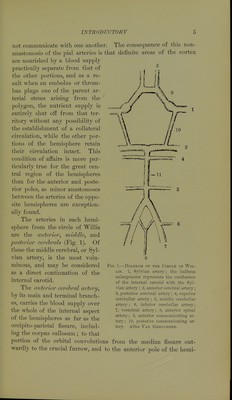
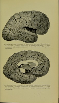
![not eonimnnicate witli one another. The consequence of this non- anastomosis of the pial arteries is that definite areas of the cortex are nourished by a blood supply practically separate from that of the other portions, and as a re- sult when an embolus or throm- bus plugs one of the j)arent ar- terial stems arising from the polygon, the nutrient supply is entirely shut off from that ter- ritory without any possibility of the establishment of a collateral circulation, while the other por- tions of the hemisphere retain their circulation intact. This condition of affairs is more par- ticularly true for the great cen- tral region of the hemispheres than for the anterior and poste- rior poles, as minor anastomoses between the arteries of the opjDO- site hemispheres are exception- ally found. The arteries in each hemi- sphere from the circle of Willis are the anterior, middle, and posterior cerebrals (Fig. 1). Of these the middle cerebral, or Syl- vian artery, is the most volu- minous, and may be considei-ed as a direct continuation of the interna] carotid. Tlie anterior cerebral artery, by its main and terminal branch- es, carries the blood supply over the whole of the internal aspect of the hemispheres as far as the occipito-parietal fissure, includ- ing the corpus callosum ; to that portion of the orbital convolutions from the median fissure out- wardly to the crucial furrow, and to the anterior pole of the hemi- FiG. 1.—Diagram of the Circle of Wil- lis. 1, Sylvian artery; the bulbous enlargement represents the confluence of the internal carotid with the Syl- vian artery ; 2, anterior cerebral artery ; 3, posterior cerebral artery; 4, superior cerebellar artery; 5, middle cerebellar artery ; 6, inferior cerebellar artery; 7, vertebral artery; 8, anterior spinal artery; 9, anterior communicating ar- tery ; 10, posterior communicating ar- tery. After Van GEnrcnTEN.](https://iiif.wellcomecollection.org/image/b21294690_0031.jp2/full/800%2C/0/default.jpg)



 No text description is available for this image
 No text description is available for this image
 No text description is available for this image