Licence: Public Domain Mark
Credit: Indurative mediastino-pericarditis / by Thomas Harris. Source: Wellcome Collection.
23/78 page 17
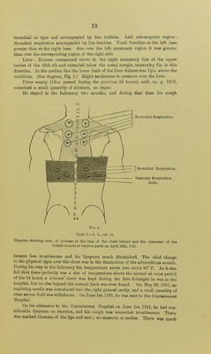
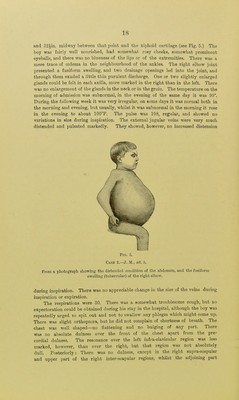
![illness (April, 1891), and heard over the inner extremity of the first inter- costal space, at the left borderof the sternum, was most probably conducted fi'om the trachea, as it may be found in this position occasionally in healthy individuals. A similar explanation applies to the bronchial respiration which was audible at the same period over the upper part of the inter- scapular region of both sides. Having regard to the appearances pre- sented at the post-mortem examination, I do not think that the bronchial breathing, heard at the posterior and upper part of the chest, could have been connected with the mediastinal affection, but that it was probably simply a pronounced example of a type of breathing which is not uncommon in this region, especially in children, who present neither an affection of the lungs nor of the mediastinum. [See also the report and comments on Case 2.] The following case is an illustration of Class II., which comprises a group of cases, where, on post-mortem examination, the pericardial cavity is found to be obliterated by adhesions, and the exterior of the pericardium is adherent to surrounding parts, whilst there is little or no increase of fibrous tissue throughout the mediastinum generally; a condition which has been termed Pericarditis interna and externa. Case 2.—Dyspncea; cyanosis; tubercular disease of the elbow joint; cardiac dilatation; engorgement of the veins of the nech; enlarged liver and ascites; very slight anasarca of the lower extremities. Repeated paracentesis of the abdomen. Bronchitis and catarrhal pneumonia, and death from gi-adual cardiac failure. [From notes by Dr. R. W. Marsden.] J. M., aged 8 years, was admitted to the Manchester Royal Infirmary, on April 7th, 1893, complaining chiefly of great distension of the abdomen. His mother gave the following account of his illness : The boy had been quite well up to 5J years of age, i.e., 2J years before he was admitted into the Infirmary. The only illness which he had had before that time was scarlet fever. At 5J years of age he was noticed to be looking ill, to be getting thinner, and to have a slight cough. At that time, however, he had no swelling of the abdomen or elsewhere. Three months later the abdomen commenced to enlarge, and after another period of three months had elapsed he was admitted to the Manchester Clinical Hospital. During the past two years the mother stated that the abdomen had been tapped 23 times, but Dr. Railton, under whose care he was, has kindly looked up the records of the'case for me, and from these we only have evidence that during the past two years the boy had been tapped 13 times. About 12 months previous to his admission into the Manchester Infirmary, signs of disease in the right elbow joint appeared, suppuration had subsequently ensued, and the joint had been drained. On admission the most prominent feature of the case was the enormous distension of the abdomen, which measured 33in. in circumference at the umbilicus B](https://iiif.wellcomecollection.org/image/b20421011_0023.jp2/full/800%2C/0/default.jpg)


 No text description is available for this image
 No text description is available for this image
 No text description is available for this image