Licence: In copyright
Credit: Pathology and treatment of diseases of women. Source: Wellcome Collection.
32/500 page 12
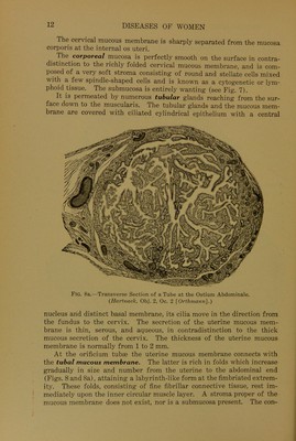
![The cervical mucous membrane is sharply separated from the mucosa corporis at the internal os uteri. The corporeal mucosa is perfectly smooth on the surface in contra- distinction to the richly folded cervical mucous membrane, and is com- posed of a very soft stroma consisting of round and stellate cells mixed \\ ith a few spindle-shaped cells and is known as a cytogenetic or lym- phoid tissue. The submucosa is entirely wanting (see Fig. 7). It is permeated by numerous tubulär glands reaching from the sur- face down to the muscularis. The tubulär glands and the mucous mem- brane are covered with ciliated cylindrical epithelium with a central Fig. 8a.—Transverse Section of a Tube at the Ostium Abdominale. (Hartnack, Obj. 2, Oc. 2 [Orthmann].) nucleus and distinct basal membrane, its cilia move in the direction from the fundus to the cervix. The secretion of the uterine mucous mem- brane is thin, serous, and aqueous, in contradistinction to the thick mucous secretion of the cervix. The thickness of the uterine mucous membrane is normally from 1 to 2 mm. At the orificium tubte the uterine mucous membrane connects with the tubal mucous membrane. The latter is rieh in folds which increase gradually in size and number from the uterine to the abdominal end (Figs. 8 and 8a), attaining a labyrinth-like form at the fimbriated extrem- ity. These folds, consisting of fine fibrillär connective tissue, rest im- mediately upon the inner circular muscle layer. A stroma proper of the mucous membrane does not exist, nor is a submucosa present. The con-](https://iiif.wellcomecollection.org/image/b28131484_0032.jp2/full/800%2C/0/default.jpg)


 No text description is available for this image
 No text description is available for this image
 No text description is available for this image