A manual of the dissection of the human body / by Luther Holden ; with notes and additions by Erskine Mason.
- Luther Holden
- Date:
- 1868
Licence: Public Domain Mark
Credit: A manual of the dissection of the human body / by Luther Holden ; with notes and additions by Erskine Mason. Source: Wellcome Collection.
Provider: This material has been provided by the Francis A. Countway Library of Medicine, through the Medical Heritage Library. The original may be consulted at the Francis A. Countway Library of Medicine, Harvard Medical School.
27/608 (page 13)
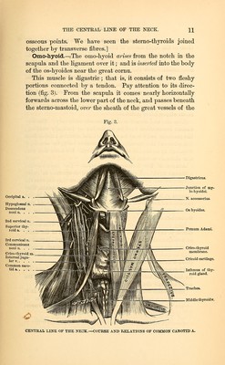
![particularly notice the strong layer of fascia •which lies under the sterno-mastoid, and forms the under part of its sheath. It is attached to the angle of the jaw, thence descends over and protects the great vessels of the neck, and is firmly con- nected to the clavicle and first rib. This fascia prevents mat- ter from coming to the surface, when suppuration takes place by the side of the pharynx. Remove this fascia, taking care not to remove with it the descendens noni and communicantes noni nerves, which cross the sheath of the common carotid. Dissect out any absorbent glands which lie about the sheaths of the great vessels. Course and Relations of the Common Carotid Artery.— The common carotid arises on the right side from the arteria innominata; on the left, from the arch of the aorta. It as- cends in front of the bodies of the cervical vertebrae, by the side of the trachea, thyroid gland, and larynx, as high as the upper border of the thyroid cartilage, and then divides into the external and internal carotid. Thus a line drawn from the sternal end of the clavicle to the angle of the jaw will nearly indicate its course. It is contained in a sheath of cervical fascia. In the same sheath are the internal jugular vein and the pneumogastric nerve. The vein lies on the outer side of, and parallel with the artery : the nerve lies behind and be- tween the artery and vein. Behind the sheath is the sympa- thetic nerve. Lastly, along the vertebral column the sheath lies successively upon the longus colli and the rectus capitis anticus major muscles. [Frequently well marked septa are found within the sheath, dividing it into three separate compartments for the vein, artery and nerve.] At the lower part of the neck the carotid artery is deeply seated:—it is covered by the sternal portion of the sterno- mastoid, the stemo-hyoid and thyroid muscles, and about two inches above the clavicle it is crossed by the omo-hyoid. Above this point the artery becomes more superficial, and is covered by the platysma, cervical fascia, and only slightly overlapped by the sterno-mastoid. Lying immediately upon the sheath of the artery we find the descendens noni and communicantes noni nerves. Above aU, observe that the sheath is crossed by three veins, namely, the facial, the supe-](https://iiif.wellcomecollection.org/image/b21059342_0027.jp2/full/800%2C/0/default.jpg)