A manual of the dissection of the human body / by Luther Holden ; with notes and additions by Erskine Mason.
- Luther Holden
- Date:
- 1868
Licence: Public Domain Mark
Credit: A manual of the dissection of the human body / by Luther Holden ; with notes and additions by Erskine Mason. Source: Wellcome Collection.
Provider: This material has been provided by the Francis A. Countway Library of Medicine, through the Medical Heritage Library. The original may be consulted at the Francis A. Countway Library of Medicine, Harvard Medical School.
28/608 (page 14)
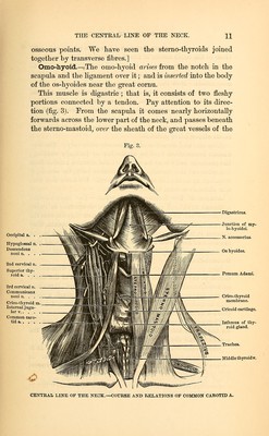
![rior, and inferior thyroid, which empty themselves into the in- ternal jugular. This is the general rule, and we draw especial attention to it, because these veins are so liable to be over- looked and injured in the operation of tying the carotid. It is plain that it is easier to tie the common carotid above the omo-hyoid than below it. We make an incision (three inches long) down the inner border of the stemo-mastoid; we cut through the platysma and cervical fascia, draw aside the overlapping edge of the sterno-mastoid, and expose the sheath of the vessel. We then make a small opening in the sheath large enough to admit the aneurismal needle, and tie the vessel, taking care not to include the pneumogastric or descen- dens noni nerves in the ligature. [To tie the artery, the head should be well thrown back, and when the operation is to be above the omo-hyoid, an incision should be made a little below the angle of the jaw to the cri- coid cartilage. When below this muscle, we should commence on a level with the cricoid cartilage. The sterno-mastoid must be well drawn to the outside before the vessel is ob- served; the descendens noni here runs on the tracheal side, and the recurrent laryngeal nerve behind it. In either opera- tion the needle should be passed from without, inwards.] In -what respects the Left Carotid differs from the Right.—In the first part of its course the left carotid differs from the right in the following particulars :— 1. It arises from the arch of the aorta, is therefore longer and deeper seated than the right, and is covered by the first bone of the sternum. 2. It is crossed by the left vena innominata. 3. It is in close relation with the oesophagus. 4. It is in close relation with the left recurrent nerve. 5. It is in close relation with the left thoracic duct. Division of the Common Carotid.—The common carotid at its division is often a little bulbous. This dilatation is in some instances so marked, that during life I have seen it mis- taken for an incipient aneurism. It is necessary to be aware that the carotid sometimes divides much lower than usual. Several times I have seen the division as low as the level of the cricoid cartilage. [The common carotid may be absent—the external and inter-](https://iiif.wellcomecollection.org/image/b21059342_0028.jp2/full/800%2C/0/default.jpg)