Principal characters of the Protoceratidae / by O.C. Marsh.
- Othniel Charles Marsh
- Date:
- 1897
Licence: Public Domain Mark
Credit: Principal characters of the Protoceratidae / by O.C. Marsh. Source: Wellcome Collection.
Provider: This material has been provided by The Royal College of Surgeons of England. The original may be consulted at The Royal College of Surgeons of England.
14/26 page 176
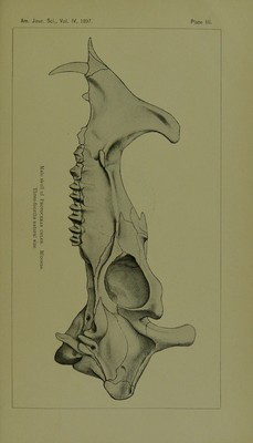
![interval between the canine and second premolar, as shown in the figure cited. The remaining upper premolars correspond closely with those of Protoceras in form, and this is true, also, of the molars. The lower incisors of Calops are small and pro- cumbent. The canine also was small, and probably'similar in form to' the incisors. The first lower premolar is caniniform in shape, with a single root, and a sharp compressed crown, which came nearly in apposition to the superior canine. The remaining lower premolars and molars agree closely except in size with those of Protoceras. The remains of Calops now known all appear to have per- tained to females, and this naturally suggests the question—what the male skull was like, and especially whether it was provided with horns. The probabilities at present are in favor of the latter view, but it must be left to future discoveries to settle that poiut. All the known remains of Protoceras and Galops are from the upper Miocene of South Dakota. The horizon, which is a definite one, has been appropriately called by Dr. Wortman the Protoceras beds. They appear to be identical with the series in Oregon which the writer had previously named the Miohippus beds, as that genus and several others are common to both regions. Tale University, New Haveu, Conn., July 24, 1897. EXPLANATION OF PLATES. Plate II. Male skull, with lower jaw, of Protoceras celer, Marsh; ohlique side view. Three-fourths natural size. Plate III. The same skull; seen from the left side. Three-fourths natural size. Plate IV. The same skull; seen from above. Three-fourths natural size. Plate V. The same skull; seen from below. Three-fourths natural size. Plate VI. Figure 1.—The same skull; seen from in front. Figure 2.—Front of skull of Protoceras comptus, Marsh; seen from below; young female, showing deciduous dentition. Both figures are three-fourths natural size. Plate VII. Figure 1.—Skull, with lower jaw. of Calops consors, MarBh; seen from the left. Figure 2.—The same skull; seen from above. Figure 3.—Natural brain cast of Protoceras celer; female; side view. Figure 4.—The same; seen from above. The figures are all one-half natural size. [To be continued.]](https://iiif.wellcomecollection.org/image/b22322747_0016.jp2/full/800%2C/0/default.jpg)