A text-book of embryology for students of medicine / by John Clement Heisler.
- Heisler, John Clement.
- Date:
- 1907
Licence: In copyright
Credit: A text-book of embryology for students of medicine / by John Clement Heisler. Source: Wellcome Collection.
Provider: This material has been provided by UCL Library Services. The original may be consulted at UCL (University College London)
437/482 page 413
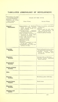
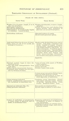
![Tabulated Chronology of Development (Continued). STAGE OF THE FETUS. Ninth Week. Third Month. Weight, 15 to 20 grams; length, 25 to 30 mm. (1 to If inches). Hard palate completed. Free tail has disappeared. Differentiation of lymph-nodes begins (0. Schultze). Cloaca divided. Weight (end of month), 4 ounces; length, 2| inches. At first chorion leve and chorion fron- dosum present; later, formation of placenta (see second frontispiece). Ppripa vc\ \ n m \ n c\ i ofi i pf] lavcllUll o \ ott 1U yJL V Coot/la. Blood-vessels penetrate spleen. Anal canal formed by division of cloaca. (Anus opens at end of 2d month, ac- cording to Tourneux.) Mouth-cavity divided from nose (end of month). Soft palate completed (11th week). Papillse of tongue. Evagina- tion for tonsil. Intestine begins to re- cede within abdomen (10th week). Ro- tuition /\c t ruri n p Vi Vt>T,mi fnfin qr-\r\p_ \.<%\j1vjLI \Ji. o HJlllcl'L/Il • V CI 111 I lvJi 111 dJJj..JCIl dix as a slender tube. Omental bursa. Gastric glands and glands and villi of intestine fairly well formed (10th week). Liver very large. Peritoneum Vine it« prlnlt Vii«tolncripo 1 r'VinrjipfprQ Epiglottis. External genitals begin to show dis- tinctions of sex. Ovary and testis distinguishable from each other. Kidney has its characteristic features. Urogenital sinus acquires its own aper- ture by division of cloaca. Union of testis with canals of Wolffian body complete. Testes in false pelvis. Ovaries descend. Prostate begun (12th week). Corium proper present as distinct layer. Nails not quite perfectly formed. T^PcriTmino' nf HpvpI nnm put O-f TlPlT £1Q JJCgllllllllg KJl. UC V t i|'lllCUl Ul ±±<A>1.L ao solid ingrowths of epithelium. Corpus striatum indicated. Corpora quadrigemina represented by two elevations on mid-brain roof. Cerebrum covers inter-brain. Fornix and corpus callosum begun. Fissure of Sylvius. Calcarine fissure. Crura cerebri. Restiform bodies. Pons. External ear indicated (Fig. 170). Ciliary processes indicated. Eyes nearly in normal position. Eyelids begin to adhere to each other. Centers of ossification of presphenoid, of lesser wings of sphenoid, and of shafts of metatarsal bones. Beginning ossification of occipital bone, of tympanic, of spine of scapula, of ossa innominata. Cartilaginous arches of vertebrae close. Limbs have definite shape ; nails almost perfectly formed.](https://iiif.wellcomecollection.org/image/b21286991_0437.jp2/full/800%2C/0/default.jpg)