A practical treatise on urinary and renal diseases, including urinary deposits / by William Roberts.
- William Roberts
- Date:
- 1872
Licence: Public Domain Mark
Credit: A practical treatise on urinary and renal diseases, including urinary deposits / by William Roberts. Source: Wellcome Collection.
529/654 (page 501)
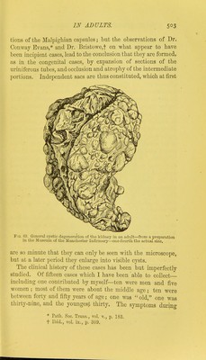
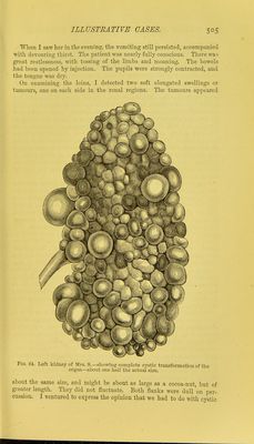
![The degeneration has not always been found in so extreme a degree as in this case of Dr. Lever. Generally, some rem- nants of secreting texture (uriniferous tubes and Malpighian tufts) have been detected in the interstices between the cysts. In some cases the external surface is smooth, while the interior presents a spongy or cavernous structure, which, under the microscope, resolves itself into myriads of minute cysts. The researches of Virchow and Forster have fully demonstrated, that the cysts in these cases are originally produced by dilata- tion of short sections of the uriniferous tubes into pouches; these pouches afterwards become enlarged and separated from each other, and at length form distinct cysts. They are lined with a tesselated epithelium, and contain, at first, a urinous fluid, which, at a later period, when the cysts attain a larger size, becomes albuminous. It is curious that malformations of the pelvis of the kidney, of the ureter, bladder, or urethra, or of some other part of the body, nearly always co-exist with congenital cystic degene- ration of the kidneys. Sometimes, however, the lower urinary passages are perfectly open. ^ Virchow first pointed out the mechanical cause of this disease. In all the cases examined by him, there was found an imperforate state (atresia) of the straight ducts which terminate on the papilla?; and he conjectures that this had arisen from intra-uterine inflammation of the ducts of the papilke, which ended in adhesion of their parietes and closure of their calibre. He further believes that the usual cause of this inflammation is the impaction of uric acid or the urates (Harnsaure-infarct) in the straight canals (see p. 469). The closure of the excretory ducts necessarily causes stagnation and accumulation of the urine throughout the entire organ, and leads to dilatations of sented the usual lobulated appearance of the foetal organs, but on section numerous small transparent cysts were found of the size of peas, in a matrix of a light grayish, colour, from which they could not be detached, and which contained a clear serous fluid. The distinction between the cortical and medullary portions was totally obliterated, but the outline of the calices could be indistinctly traced. The ureters were pervious ; bladder empty. The right kidney, even after exposure to the air for some days, weighed 6 oz • the left was ap]>arcntly of equal size.](https://iiif.wellcomecollection.org/image/b20403963_0529.jp2/full/800%2C/0/default.jpg)