Handbook of diseases of the skin : illustrated.
- Ziemssen, H. von (Hugo), 1829-1902.
- Date:
- 1885
Licence: Public Domain Mark
Credit: Handbook of diseases of the skin : illustrated. Source: Wellcome Collection.
Provider: This material has been provided by the Gerstein Science Information Centre at the University of Toronto, through the Medical Heritage Library. The original may be consulted at the Gerstein Science Information Centre, University of Toronto.
34/686 (page 16)
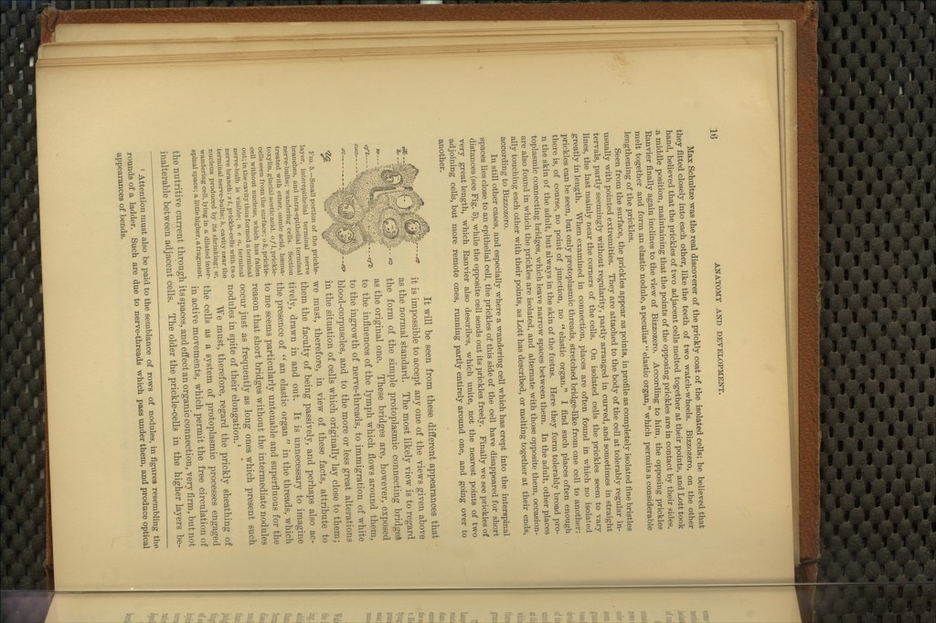
![Max Schultze was the real discoverer of the prickly coat of the isolated cells; he believed that they fitted closely into each other, like the teeth of two watch-wheels. Bizzozero, on the other hand, believed that the prickles of two adjacent ceUs melted together at their points, and Lott took a middle position, maintaining that the points of the opposing prickles are in contact by their sides. Eanvier finally again inclines to the view of Bizzozero. According to him, the opposing prickles melt together and form an elastic nodule, a peculiar elastic organ, which permits a considerable lengthening of the prickles. Seen from the surface, the prickles appear as points, in profile as completely isolated fine bristles usually with pointed extremities. They are attached to the body of the cell at tolerably regular in- tervals, partly seemingly without regularity, partly arranged in curved, and sometimes in straight lines, the last mainly near the corners of the cells. On isolated cells the prickles seem to vary greatly in length. When examined in connection, places are often found in which no isolated prickles can be seen, but only protoplasmic threads, stretched bridge-like from one cell to another; there is, of course, no point of junction, no elastic organ. I find such places often enough n the skin of the adult, but always in the skin of the foetus. Here they form tolerably broad pro- toplasmic connecting bridges, which leave narrow spaces between them. In the adult, other places are also found in which the prickles are isolated, and alternate with those opposite them, occasion- ally touching each other with their points, as Lott has described, or melting together at their ends, according to Bizzozero. In still other cases, and especially where a wandering cell which has crept into the interspinal Bpaces lies close to an epithelial cell, the prickles of this side of the cell have disappeared for short distances (see Fig. 5), while the opposite cell sends out its prickles freely. Finally we see prickles of very great length, which Ranvier also describes, which unite, not the nearest points of two adjoining cells, but more remote ones, running partly entirely around one, and going over to another. oft-.- It will be seen from these different appearances that it is impossible to accept any one of the views given above as the normal standard. The most likely view is to regard the form of the simple protoplasmic connecting bridges as the original one. These bridges are, however, exposed to the influences of the lymph which flows around them, to the ingrowth of nerve-threads, to immigration of white blood-corpuscles, and to the more or less great alterations in the situation of cells which originally lay close to them; we must, therefore, in view of these facts, attribute to them the faculty of being passively, and perhaps also ac- tively, drawn in and out. It is unnecessary to imagine the presence of '*^ an elastic organ in the threads, which to me seems particularly untenable and superfluous for the ceu without nucleus, which has fallen j-easou that short bridges without the intermediate nodules out; in the cavity thus formed a terminal . , « Ti i i • i - i nerve-bulb is visible; n e a, terminal occur ]ust as frequently as long oncs which present such nerve branch; s t, prickle-cells with two ^oduleS in Spitc of their elongation.' terminal nerve-bulbs;/i, cavity near the , .1 » j xi. • ii -u j-i, • £ nucleus produced by its shrinking; w. We must, therefore, regard the prickly sheathing of wandering cell, lying in a dilated inter- ^^^ ggjjg q,q g^ system of protoplasmic prOCCSSeS engaged spinal space; a uttiehigher,afragment. .^ ^^^.^^ movements, which permit the free circulation of the nutritive current through its spaces, and effect an organic connection, very firm, but not inalterable between adjacent cells. The older the prickle-cells in the higher layers be- «es- Fig. 5.—Small portion of the prickle- layer, interepithelial terminal nerve branches, and intra-epithelial terminal nerve-bulbs; wandering cells. Section treated with ether, osmic acid, heema- toxylin, glacial acetic acid, of I, prickle- cells seen from the surface; o fc, prickle- ' Attention must also be paid to the semblance of rows of nodules, in figures resembling tlie rounds of a ladder. Such are due to nerve-threads which pass under them, and produce optical appearances of bends.](https://iiif.wellcomecollection.org/image/b20997292_0034.jp2/full/800%2C/0/default.jpg)