Anatomia uteri humani gravidi tabulis illustrata / auctore Gulielmo Hunter = The anatomy of the human gravid uterus exhibited in figures / by William Hunter.
- William Hunter
- Date:
- [between 1842 and 1852]
Licence: Public Domain Mark
Credit: Anatomia uteri humani gravidi tabulis illustrata / auctore Gulielmo Hunter = The anatomy of the human gravid uterus exhibited in figures / by William Hunter. Source: Wellcome Collection.
21/160 (page 7)
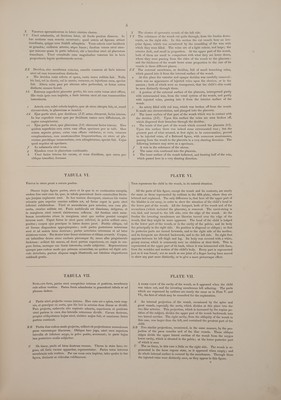
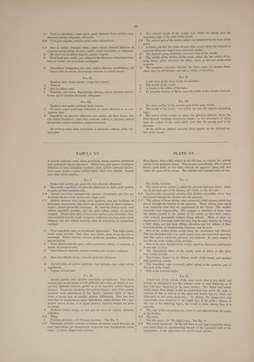
![D Vertebrarum lumborum canalis, in quo reliquia equine, obscure licet, conspiciuntur. EE Musculorum lumbos extendentium partes inferiores. F — Lumborum membrana adiposa et cutis. GGG Sacri ossa tria superiora. quedam caude HH Sacri ossa duo inferiora, primumque coccygis, firmiter coalita. ii Coccygis os secundum, quod, ope mediz cartilaginis, ad primum facile movetur. K ~—Coceygis ossa duo inferiora in unum coalita, parique ad secundum mobilitate praedita, L Sacri spina. M Sacri canalis. N Musculorum abdominis et integumentorum anguli inferioris pars, in ossis ilium spinam reflexa. Vena inguinalis magna epigastricam emittens, quam etiam arteria ejusdem nominis comitatur. P Femoris musculi, ex osse pubis orti, quique in osse eo sejungendo fuerant perscissi. Q Os pubis supra foramen magnum disscissum. # Idem os, ubi ramo parvo ischii committitur, disscissum. O In loco quo litera P occurrit, lineis punctis adumbrata representatur partis ejus ossis pubis, quee abscissa fuerat, figura. Nota * iisdem lineis adumbratam symphysin ostendit. S Pars carnosa anum inter et os coccygis. YT Anus omnino apertus. U __ Recti pars inferior hac sectione exposita. In hac figura videas, intes- tinum idem ab ano sursum progrediens oculo dum persequeris, id se retrorsum deflexisse, nempe ut ad faciem internam ossis coccygis ve- niret. V Hic rectum ad latus sinistrum se deflexit, solamque membranam suam cellulosam in hac sectione cernere licebat. WW Rectum sectione apertum, ubi ante coccygis os primum, sacrique ossa tria inferiora, decurrebat. X Hic rectum omniuo in latere dextro jacebat, nec id aperuit scissura. Y Rectum disscissum, ubi de latere dextro ad os secundum sacri decurre- bat, et in flexuram coli sigmoideam cursum suum tenebat. Z_ Vena cava. Arteria iliaca dextra. In hoc loco vena iliaca sinistra fuerat disscissa; cera autem, que injecta fuerat, utpote friata, jam delapsa, vena haud satis clare se in conspectum dedit. Perinzi sectio. Labii dextri, ad partem inferiorem, facies interna. Nymphe dextre, ad partem inferiorem, facies interna. Coarctatio, hymenisve reliquiz, ubi incipit vagina. Meatus urinarii extremitas. Vagine cavum. In hoc loco vagina et rectum sunt coalita. i] ~ Sp SSS. oy No Rectum vagina densius. = Vagine pars anterior, cum urethra et vesica urinaria conjuncta. Zi Os uteri in vagina, ad os coccygis vergens. Quamvis in hoc cadavere fundus uteri dextrorsum inclinatus fuerat, os uteri tamen adeo ad dextrum se tenebat, ut cultrum anatomicum has partes secantem fugeret. mm Ad os tince, uteri et vaginz substantia coalita. nn Uteri in duas partes zequales secti margo posterior. oo Uteri secti pars anterior. Sectio hec nequaquam per medium uteri se habebat, sed ad latus dextrum haud paululum; etenim ut figure pre- cedentes commode possent delineari, cadaver hoc niodo necesse habui disponere. p Os uteri intus. gq Uteri paries internus membranis suis vestitus. Fundus uteri in hac figura non representatur; tum quoniam id a proposita ratione alienum duxi, tum guoniam uterus adeo flaccidus et tener tunc temporis erat, ut in situ suo naturali, ad arbitrium et usum pictoris, minime potuerit retineri. Membrane, tempore quo tabula hec delineabatur, ipsze se ab utero fere prorsus sejunxerant. In parte superiori, decidua, ubi se ab utero re- ceperat, venulis abundavit ; juxta os uteri, vix ullum horum vasorum vestigium apparuit. y Meatus urine. Vesice urinariz pars inferior, uteri cervicem inter et vagine partem superiorem sita. Omnis vesice portio post symphysin pubis situ jamdu- dum abscissa fuerat. Melius autem mihi visum est, figuram vel curtam dare, quam aliquod ingenio excogitatum pingere. ~! D ‘The canal of the lumbar vertebra, in which some remains of the cauda equina are indistinctly seen. EF The lower part of the extending muscles of the loins. Ff The adipose membrane and skin of the loins. GGG The three superior component bones of the os sacrum. HH The two lowermost bones of the sacrum, and the first of the coccyx, firmly anchylosed. I The second bone of the coccyx, movable on the first by means of an intermediate cartilage. K The two last bones of the coccyx grown into one, and movable on the second bone by the intervention of a cartilage. L ‘The spine of the sacrum. M _ The spinal canal in the sacrum. N Part of the lower flap of the abdominal muscles and integuments turned over the spine of the os ilium. O The great inguinal vein, sending off the epigastric, which is accom- panied with the artery of the same name. The muscles of the thigh which arose from the os pubis, and which were cut through when that bone was removed. The os pubis cut through above the foramen magnum. The same bone cut through at its conjunction with the small branch of the ischium. Where the letter P stands, is represented, in dotted outlines, the figure of that part of the os pubis which was cut off, and the mark * is upon the symphysis in the same outlines. S The fleshy part between the os coccygis and the anus. T The anus considerably opened. U The lower part of the rectum laid open by the section. In tracing the gut from the anus upwards, we see, from this figure, that it takes a bend backwards, to get at the inside of the os coccygis. Oe) 0 VY Here the rectum made a turn to the left side, and its surrounding cel- lular membrane only was seen in the section. WW The rectum laid open by the section, where it runs before the first bone of the coccyx, and the three lowermost pieces of the sacrum. X Here the rectum lay entirely in the right side, and was not opened by the section. Y The rectum cut through by the section, where it passed from the right side across the second bone of the sacrum, and was continued into the sigmoid flexure of the colon. Z The vena cava. a The right iliac artery. b Here the left iliac vein was cut through; but the brittle wax with which it was filled had fallen out, and the vein was seen indistinctly. e The section of the perineum. d The inside of the lower part of the right labium. e The inside of the lower part of the right nympha. f The stricture at the beginning of the vagina, or remains of the hymen. g The extremity of the meatus urinarius. hh ‘The cavity of the vagina. 2. The compound substance of the vagina and rectum, the latter of which is considerably the thickest. & The forepart of the vagina united with the urethra and bladder. ‘1 ‘The mouth of the womb in the vagina, directed towards the os coc- cygis. Though the bottom of the womb, in this case, was directed towards the right side, its mouth lay so much on the right side that it was not touched in making the middle section of all the parts. The substance of the womb and vagina blended at the os tince. nn The edge of the bisected womb backwards. 00 The edge of the womb forwards. This section was not in the middle, but considerably on the right side. It had been made to prepare the subject for some of the preceding figures. mim p The mouth of the womb internally. q The inside of the womb lined with the membranes. The bottom of the womb is not represented in this figure; both be- cause it was not very material here, and because it was become so flaccid and tender that it could not be well kept out, in its natural situation, before the painter. The membranes were almost entirely separated from the womb, of themselves, when this figure was made. In the upper part the decidua was full of small veins, even where it parted of itself from the womb; but near the mouth of the womb hardly any such vessels appeared. y The meatus urine. s The lower part of the bladder, placed between the neck of the womb and the upper part of the vagina. The upper part of the bladder, which was situated behind the symphysis of the pubes, had been cut away be- fore this section was made; and we chose to leave the figure imperfect rather than venture to delineate from fancy.](https://iiif.wellcomecollection.org/image/b32882142_0021.jp2/full/800%2C/0/default.jpg)