Anatomia uteri humani gravidi tabulis illustrata / auctore Gulielmo Hunter = The anatomy of the human gravid uterus exhibited in figures / by William Hunter.
- William Hunter
- Date:
- [between 1842 and 1852]
Licence: Public Domain Mark
Credit: Anatomia uteri humani gravidi tabulis illustrata / auctore Gulielmo Hunter = The anatomy of the human gravid uterus exhibited in figures / by William Hunter. Source: Wellcome Collection.
28/160 (page 14)
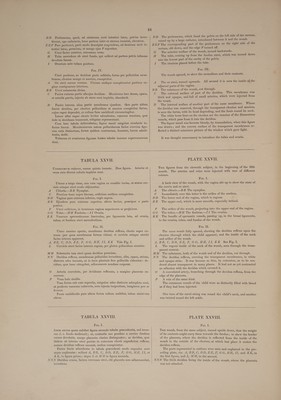
![Litere sequentes ad figuram magis elaboratam spectant. XX Totius vagine substantia ex uteri cervice et vesica urinaria excisa et in rectum devoluta, ut os uteri in conspectum prodiret, Y —_ Oris uteri labium anterius. Z ~~ Bjusdem labium posterius. Vagina ad hunc modum aperta, os uteri, nimirum adminiculo suo jam a latere sublato, dextrorsum propendebat. TABULA XXIII. Ocravum cadaver, sexto gestationis mense. Uteri ac membranarum parte anteriori sublata, exhibetur foetus cum parte placentz et funiculi umbilicalis. Uteri vasa cera impleta fuerant. A Vesica urinaria, respectu habito ad uterum, in situ suo naturali; modice distenditur, et ramis quibusdam majoribus venarum hypogastri- carum circumtegitur. B Vagine partis postice facies interna. CC Vasa hypogastrica ad cervicem uteri decurrentia et ramos ad vesicam urinariam et vaginam transmittentia. DD _ Vasaspermatica, duplicationem ligamenti lati intrantia. EE Tube.—F F Fimbrie. GG Ligamenti lati lamella posterior: anterior cultro anatomico sublata fuerat, ut vasa spermatica, ad fundum uteri ascendentia, oculo plenius occurrerent. H H Ligamenta rotunda. In sinistro, arteria preecipua, convoluta, a sper- matica descendens conspicitur. TIT Uteri substantiz totius et membranarum sectio, qua uteri et secun- darum paries anterior, ad eorum contenta exhibenda, sublatus fuerat. K Funis umbilicalis, prope locum ubi in placenta desinit. Placenta uteri parti postice, versus fundum, adherebat. Funis umbi- licalis, primo deorsum, super humerum sinistrum, deinde sursum, pone corpus infantis, ad finem suum in placenta transibat. TABULA XXIV. AB eodem cadavere. Fria. I. Placenta uteri fundo et parti posticee adhzrescens ; substantia ejus spongiosa, injecta per vasa uterina cera, turgescit. Pleraeque partes lineis adumbratz, in tabula praecedenti ad plenum sunt exposite. A Vesica urinaria. —B Vagina. CC Vasa hypogastrica.—D D Vasa spermatica. EE Tube.—/F SF Fimbrie. GG Ligamenta lataa—H H Ligamenta rotunda. III Uteri et membranarum sectio.— K Funis umbilicalis. i. Placenta utero adheerescens. Nulla pars cere, in vasa uterina injectae, ad ramos vasorum illorum que funem umbilicalem conficiunt, transierat ; vasa illa sanguinem so- lummodo continentia, obscure designata erant, ubi ex fune umbilicali in faciem internam placentz se immittebant. Cellule autem in placente parte spongiosa, omnes eodem modo cera, vel coerulea in venas uteri in- jecta, vel rubra in arterias infusa, turgescebant. Hance rem indicat figura secunda, M Membrane ex margine placente exeuntes, et uterum undique in- vestientes. Placente margo hic paulo elatior est, et magis conspicuus, substantia ejus spongiosa cera distensa. Fre. II. Portio aliqua placente transverse secta, ut substantia ejus spongiosa appareret, ejusdemque cera adimplete crassitudo. Aacentm@m s srfiejes j . ] A _ Placent superficies interna.—B Placente superficies externa. Y . we ar oT A any C Me mbranat um portiuncula, ex margine placenta, cera injecta turges- centis, et in figuram rotundiorem adaucte, exiens. ™ we . oy aes ey “7 >) as ac +4 5 1 Cere coerulex, primum per venas uteri Injectee, maxima pars ad super- ficiem ejus internam propulsa fuerat ; ceraque rubra, postea per arterias injecta, in partibus exterioribus restabat ; per totam autem ejus substan- tiam colores hi duo plus minusve commixti sunt. The following letters relate only to the more finished figure, viz. XX The whole substance of the vagina, in the right side, cut from the neck of the womb and bladder, and turned down over the rectum, to shew the orifice of the womb. Y The anterior lip of the orifice. Z_ The posterior lip of the same. - When the vagina was thus opened, the lateral support being removed, the os uteri pushed out towards the right side. PLATE XXIII. From the eighth subject, at six months. A fore-view of the womb, which was injected; the anterior part, both of the womb and of the membranes, having been cut away, and the liquor amnii taken out to shew the foetus, with a part of the placenta and of the navel-string. A The bladder, in its situation with respect to the womb. It is mode- rately distended, and is covered with some large branches of the hypo- gastric veins. B The inside of the posterior part of the vagina. CC The hypogastric vessels, going into the neck of the womb, and sending branches to the bladder and vagina. DD The spermatic vessels, going into the duplicature of the broad ligament. EE The tubes.—/ Ff The fimbriz. GG The posterior lamella of the broad ligament: the anterior had been removed by dissection, to give a clearer view of the spermatic vessels, in their ascent to the fundus of the womb. HH The round ligaments. In the left is seen a large convoluted artery, coming down from the spermatic. III The section of the whole substance of the womb, and of the mem- branes, by which the fore-part of the womb and of the secundines was removed, to expose their contents. K The navel-string, near its termination in the placenta. The placenta adhered to the posterior part of the womb, towards the fundus. The navel-string passed first downwards, over the left shoulder, and then upwards, behind the body of the child, to its termination at the placenta. PLATE XXIV. From the same subject. Fig. I. The placenta, adhering to the fundus and back-part of the womb : its spongy substance is filled by the injection of the uterine vessels. Most of the parts in outlines were more fully represented and explained in the preceding plate. A The urinary bladder.—B The vagina. CC The hypogastric vessels. —D D The spermatic vessels. EE The tubes.—/'F The fimbriz. GG _ The broad ligaments.—H H The round ligaments. III The section of the womb and membranes.—K The navel-string. LL The placenta, adhering to the womb. None of the wax, injected into the vessels of the womb, had passed into the branches of those vessels which compose the navel-string ; and as they contained only some blood, they were not distinctly marked, where they spread, from the navel-string, over the internal surface of the placenta. But the cells, or interstices in the spongy part of the placenta, were universally loaded with wax; either the blue, which was injected into the veins of the womb, or the red, which was thrown into the arteries. This is illustrated by Fig. II. M The membranes, coming out from the edge of the placenta, and in- vesting the womb all around. The edge of the placenta, in this case, was much more elevated and distinct, its spongy substance being distended. rg lle A section of half of the placenta, principally to shew what thickness it had acquired, by its spongy cavities being filled with wax. A Its internal surface——B Its external surface. C A small portion of the membranes, going off from the edge of the pla- centa, which was thickened, and rounded, by the injected wax. Most of the blue wax, which was first injected by the veins of the womb, was driven on towards the internal surface ; and the red wax, which was afterwards injected by the arteries, was lodged principally in the outer parts; but the two colours were, more or less, blended through](https://iiif.wellcomecollection.org/image/b32882142_0028.jp2/full/800%2C/0/default.jpg)