Principles of human physiology / by William B. Carpenter.
- William Benjamin Carpenter
- Date:
- 1869
Licence: Public Domain Mark
Credit: Principles of human physiology / by William B. Carpenter. Source: Wellcome Collection.
Provider: This material has been provided by Royal College of Physicians, London. The original may be consulted at Royal College of Physicians, London.
80/1110 page 46
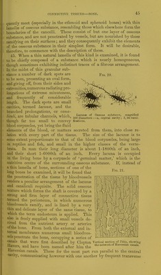
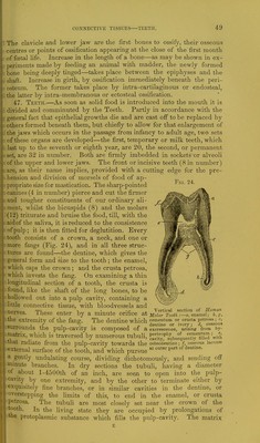
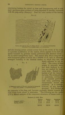
![CONNECTIVE TISSUES—BONE. branches, so that the whole system forms an irregular network, solid texture, and adapted for the Fig. 22. pervading every part of the establishment of vascular communication throughout. The dia- meter of the Haversian canals varies from l-2500th to l-200th of an inch or more; their average diameter may be stated at about 1-500th of an inch. The Arteries and Veins usually occupy sepa- rate channels, and those inclosing the latter are, in some instances, a in the diploe of the flat bones of the skull, of extraordinary ampli- tude. When a transverse section of a long bone is made, the open orifices of the longitudinal canals present themselves at intervals, sometimes con- nected by a transverse canal where the section happens to traverse this. Around these orifices the osseous matter is arranged in the form of cylin- drical lamella?, producing the appearance of concentric circles, the number varying from five to twenty for each Haversian canal. In the spaces intervening between the lamella?, numerous la- Minute structure ot Bone, an shown in a thin section cut ., j , transversely to the direction of the Haversian canals :— CUna? are Situated, trie cana- 1, one of the Haversian canals surrounded by its con- Ijculi from which penetrate centric lamellae; the lacunae are seen between the lamellae, _. . . ^ but the radiating tubuli are omitted; 2, an Haversian the adjoining lamella?, pro canal with its concentrio laminae, lacunae, and radiating ,],,,' „a fir Ct^o-rrxatr* lio tubuli; 3, the area of one of the canals; 4, 4, intervening QUClnS) as UT- OAarpey na lamellae; between these lamellae at the upper part of the stated, the same appearan figure, several very long lacunae with their tubuli are seen. •, •. In the lower part of the figure the outlines of two other as WOUld be Seen On DOrin canals are given, in order to show their form and mode of JjoleS to some depth in arrangement in the entire bone. , f . straight or crooked directio through the leaves of a book, excepting only that the passages hav proper parietes. On minute examination of bones softened in acid, th lamella? are found- to present a well marked fibrous structure, the fibres) being transparent and decussating each other in the form of an extremeljj fine network, and they are often connected or bolted together by perA forating bundles of fibres of white fibrous or of yellow elastic tissuei The spongy flat bones contain from 12 to 30 per cent, of water, tha compact tissue from 3 to 7 per cent. The chemical analysis of drier! bone shows that it consists of 33 per cent, of animal matter, which, orj boiling, yields gelatine, and 67 per cent, of mineral matter, of which 5« parts are composed of phosphate of lime, 8 parts of carbonate of lime! 1 part of fluoride of calcium, and 1 part of phosphate of magnesia! The degree of hardness of bone does not altogether depend, as showil by the experiments of Dr. Stark,f on the proportion of mineral deposit they may contain; for the flexible, semi-transparent, easily-divide(I bones of fish contain as large a proportion of earthy matter as the ivory! like leg bones of the deer or sheep. As a general ride, the bones of th#j * tt Introduction to Quain's Anatomy, 7th edition, p. xcvi. Edinburgh Medical and Surgical Journal, April, 1845.](https://iiif.wellcomecollection.org/image/b24757007_0080.jp2/full/800%2C/0/default.jpg)
 No text description is available for this image
 No text description is available for this image
 No text description is available for this image