A clinical, pathological, and experimental study of fracture of the lower end of the radius : with displacement of the carpal fragment toward the flexor or anterior surface of the wrist / by John B. Roberts.
- John Bingham Roberts
- Date:
- 1897
Licence: Public Domain Mark
Credit: A clinical, pathological, and experimental study of fracture of the lower end of the radius : with displacement of the carpal fragment toward the flexor or anterior surface of the wrist / by John B. Roberts. Source: Wellcome Collection.
Provider: This material has been provided by UCL Library Services. The original may be consulted at UCL (University College London)
78/96 page 66
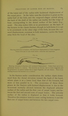
![ulna is seen to have been the seat of a fracture, though it is now united to the head of the bone. The force which carries the hand at the time of fracture away from the ulna may cause the styloid process of that bone to tear through the skin. This was probably the causation of the wound mentioned in the report of Case XIV. In Case IX. there was, in addition to the fracture of the base of the radius, which was transverse and open, an oblique fracture of the ulna an inch higher than the radial fracture. The latter was situ- ated about three-quarters of an inch from the carpal end of the bone. The articulating surface of the radius cannot be made out by palpation in fracture with forward displacement, as in dislocation forward of the carpal bones. Its plane in fractures is carried forward. This would cause the hand to assume the appearance of flexion of the wrist-joint if it were not that the extensor muscles have a tendency to cause extension between it and the first row of carpal bones and between the two rows of carpal bones, and thus bring the hand into a plane corresponding ap- proximately with that of the forearm. This condition is par- ticularly noticeable in the specimen belonging to the New York Hospital (No. 22). The way in which the articular surface looks obliquely forward is beautifully shown in the specimen from the Royal College of Surgeons of Edinburgh (No. 4). Fig. 32. Diagram of displacement on ulnar side. Specimen 22 [New York Hospital]. No lateral motion is to be expected between the carpal bones and the lower end of the radius, since the lateral ligaments,](https://iiif.wellcomecollection.org/image/b21290477_0078.jp2/full/800%2C/0/default.jpg)