Volume 1
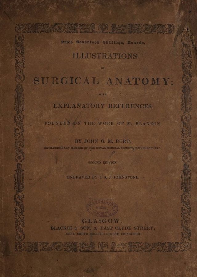
Illustrations of surgical anatomy, with explanatory references; founded on the work of M. Blandin / by John G.M. Burt ... Engraved under the direction of the Editor, by Messrs. James & John Johnstone.
- Burt, John G. M. (John Graham Macdonald), -1868.
- Date:
- [1833]
Licence: Public Domain Mark
Credit: Illustrations of surgical anatomy, with explanatory references; founded on the work of M. Blandin / by John G.M. Burt ... Engraved under the direction of the Editor, by Messrs. James & John Johnstone. Source: Wellcome Collection.
14/62
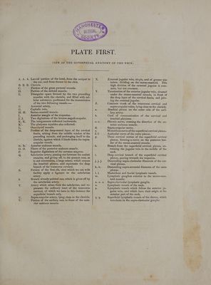
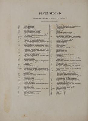
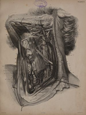
![SAHOO > 2 BRASS © aa. n q M. Portion of the ear. Portion of the cheek. Chin drawn upwards. Portion of the occiput. Superior extremity of the sternum. Right clavicular region elevated. Left clavicle directed horizontally. Right side of the neck, on which we see in profile the relative situations of the different parts. The integuments. Subcutaneous cellular substance. Layer formed above by the platysma myoides, and below by the superficial cervical fascia. Place where the cervical fascia, in the upper part single, divides into two principal layers. Anterior and posterior layers of the cervical fascia. Inferior extremity of the sterno-mastoid muscle, situated between the two layers of the cervical fascia. The anterior external jugular vein, slightly de- veloped, and confined between the two layers of the cervical fascia. Ganglia and lymphatics situated between the layers of the cervical fascia. Fatty cushion lying below the upper part of the platysma myoides. Sterno-hyoid muscle. Sterno-thyroid muscle. Left side of the neck, where the different organs have been completely laid bare, and are seen nearly in front. Hyoid bone. Inferior maxillary bone. The skin reflected backwards. The platysma myoides also reflected backwards. Hook applied to the sterno-mastoid muscle, and drawn outwards, to display the parts naturally concealed. Omo-hyoid muscle. __ Sterno-hyoid and thyroid muscles cut across. Their inferior extremities. Crico-thyroid muscle. Thyro-hyoid muscle. Anterior belly of the digastric muscle, Posterior belly cf the same muscle. Pulley of the muscle. Fibrous lamina, which proceeds frem the tendon of the muscle. Stylo-hyoid muscle. Stylo-glessus muscle. Portion of the inferior constrictor muscle of the pharynx. Portion of the hyo-glossus muscle. Mylo-hyoid muscle. Portion of the masseter muscle, Thyro-hyoid space. Por fF wre ox mm ON = ® ; 15.15.15. 16. Die 18. 19. 20. Thyroid cartilage. Crico-thyroid space, filled by an elastic membrane, exhibiting small vascular openings. Cricoid cartilage. Thyroid body. Trachea. CEsophagus inclining to the left. Portion of the brachio cephalic trunk. Left common carotid artery. Inferior thyroid artery, passing transversely be- hind the common carotid, and in front of the commencement of the cesophagus, Division of the common carotid. External carotid artery. Internal carotid artery. Superior thyroid artery. Crico-thyroid artery. Fascial artery. Sub-mental branch of the fascial artery. Lingual artery. Internal jugular vein. Anterior jugular vein. Fascial vein. Temporal vein, leaving the parotid. Lingual vein. Lateral thyroid vein. Filaments from the cervico-fascial branch of the fascial nerve. Mylo-hyoidean filament of the inferior dental and spinal nerves. Hypo-glossal, or ninth pair of cerebral nerves. - Its descending branch. Arch formed by the preceding nerve, and the de- scending internal branch of the cervical plexus. Sterno-hyoidean filament of the hypo-glossal nerve. Pneumo-gastric nerve. Superior laryngeal nerve of the left pneumo-gas- tric, dividing itself into two branches. Cardiac branch of the pneumo-gastric nerve. Inferior laryngeal or left recurrent nerve, placed in front of the cesophagus. Cervical portion of the great sympathetic. Termination of the superior cervical ganglion. Middle cervical ganglion, placed on the inferior curvature of the thyroid artery. Communicating filament of the great sympathetic with the cervical nerves. Cardiac nerve, superficial at its origin. Origin of the middle cardiac nerve. Lymphatic ganglia. Parotidean lymphatic vessels. Mastoidean do. Fascia] do. Sub-mental do. Cervical do.](https://iiif.wellcomecollection.org/image/b22011328_0001_0014.jp2/full/800%2C/0/default.jpg)
 No text description is available for this image
 No text description is available for this image
 No text description is available for this image