Autoplastic ovarian transplantation and its clinical significance / by J.H. Nattrass.
- Nattrass, John Hodgson.
- Date:
- 1910
Licence: In copyright
Credit: Autoplastic ovarian transplantation and its clinical significance / by J.H. Nattrass. Source: Wellcome Collection.
Provider: This material has been provided by The Royal College of Surgeons of England. The original may be consulted at The Royal College of Surgeons of England.
17/33 (page 13)
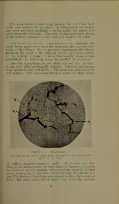
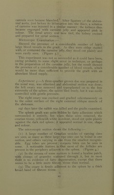
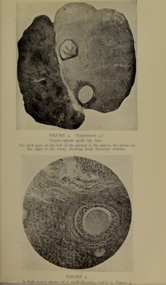
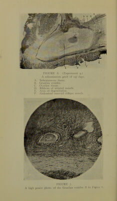
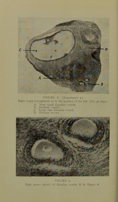
!['I he ovary j^Taftcd on tlio yiutcr surface of the external ol)li(|ue muscle had become <liv'ided into three j>arts (probably by my ligatures) ; they were granular on the surface, yellowish white in colour, and felt firm. (Figure 6.) Under the micro.scope :— (1) (Iraafian vesicles large and small may be seen. (2) Fibrous adhesion to adjacent tissue is strongly devel- oped. A peculiar feature is, that rihbon-sha]jed strands of skeletal muscle fibre seem to surround the circumference of the ovary; the spindle shape of the muscle fibre is beautifidly seen, the rest of the muscle being cut transversely. (Figure 6.) (3) The central part of tive ovary has not yet completely recovered from the condition of fatty degeneration; it pre- sents now a hyaline appearance, with large round-looking cells scattered through it, the cells being exceedingly granular. The hyaline ])art stains very feebly; it is not nearly .so exten- sive in area, as the area of fatty degeneration in the previous subcutaneous graft. (Exi)eriment I.\.) The jioints of interest in this ex])eriment are:—'I'hat the ovaries have .survived for T97 days after transplantation, and that in their new location they have still continued to develop and mature their Graafian vesicles. The remains of fatty de- generation are less in the s]denic than in the subcutaneous graft, probal)ly because the s])leen is much more vascular than the latter tissue. The degenerated area in the sul)cutaneous graft is much smaller than in a similar graft of less duration, showing that absorption and rej)air have been pnjceeding rapidly. 5.—Abdominal section was ])erformed on a half- grown doe. The left ovary was excised and jdaced in normal saline at 98.4 deg. F. The right ovary was then removed and ])laced in the exact j^osition of the left, being fi.xed to the peri- toneum and underlying muscle. The left ovary was then placed in the position of the right in a similar manner; in doing this great care was taken not to injure the tubes nor disturb their anatomical arrangement. The ovarian arteries were, tied about one inch from the ovaries. .After 97 days the rabbit was killed and tbe ovaries exam- ined. I’owel is adherent to the situation of each. The ovaries have been absorbed to such an extent that it is difficult to say if any exists at all on the right side, but the left can be felt, although very small. Microsco])ic Section shows;— 'Phe i)resence of ovarian tissue on each side, and each is crowded with Graafian vesicles, large and small. .A noticeable](https://iiif.wellcomecollection.org/image/b22428884_0017.jp2/full/800%2C/0/default.jpg)