Autoplastic ovarian transplantation and its clinical significance / by J.H. Nattrass.
- Nattrass, John Hodgson.
- Date:
- 1910
Licence: In copyright
Credit: Autoplastic ovarian transplantation and its clinical significance / by J.H. Nattrass. Source: Wellcome Collection.
Provider: This material has been provided by The Royal College of Surgeons of England. The original may be consulted at The Royal College of Surgeons of England.
9/33 (page 5)
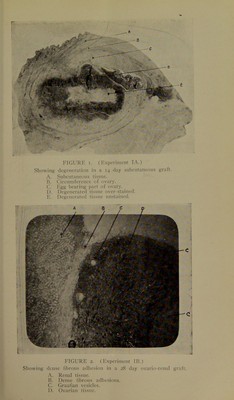
![and then curves around the anterior end, becoming attached to it by its funnel shaped ostium ; each ovary is well supplied with blood by the ovarian branch of the abdominal aorta, which can always be seen pulsating, where it crosses the lumbar muscles, transversely towards it. The low power of the microscope shows the ovary to be fairly regular in structure, the greater part of it being com- posed of fibrous stroma rich in spindle shaped cells, blood vessels and nerves. The cortical part has a large number of Graafian follicles present in it, their arrangement in parts looking not unlike a string of beads in the peripheral tissue; the follicles vary much in size, the larger ones being deeper in the stroma than the average ones. Very large ones are sometimes seen ready to burst superficially. In pregnant animals corpora lutea can always be demonstrated. With the high power the germinal epithelium and egg tubes can be made out, and the various parts of the Graafian fol- licles, i.e., the germinal vesicle with the germinal spot, the vitellus, the zona pellucida, the discus ])roligerus, the mem- brana granulosa and the surrounding theca folliculi are all easily seen. THE AUTOPLASTIC TRANSPLANTATION OF OVARIES. Experiment /.*—The abdomen of a three-quarter grown doe was clipped free from hair, washed and cleansed with per- chloride, ether was given on an open mask and abdominal section performed. The left ovarian artery was tied and the left ovary excised and placed in normal saline; a small incision was then made, through the peritoneal and capsular covering of the left kidney; a small part of the capsule was freed from the renal tissue; the ovary was then passed through the incision under the capsule and was thus retained in close irroximity to the renal tissue; the incision was then sewn,up with fine silk. The right ovary was excised in a similar manner, and the skin being well retracted from the edge of the abdominal inci- sion, it was sewn on to the outei surface of the right external oblique muscle. The abdominal wound was then closed with silk-worm gut and the rabbit placed in an artificially heated hutch. A.—Fourteen days later under ether the subcutaneous graft was removed:—The ovary was well defined and felt (|uite firm, more white in colour and seemed to be slightly larger *Thc.sc experiments were carried out 1)etween December yth, igo8, and August i6th, 1909.](https://iiif.wellcomecollection.org/image/b22428884_0009.jp2/full/800%2C/0/default.jpg)