Subcutaneous calcinosis or multiple calcification in the subcutaneous tissue / by F. Parkes Weber.
- Frederick Parkes Weber
- Date:
- 1914
Licence: In copyright
Credit: Subcutaneous calcinosis or multiple calcification in the subcutaneous tissue / by F. Parkes Weber. Source: Wellcome Collection.
Provider: This material has been provided by The Royal College of Surgeons of England. The original may be consulted at The Royal College of Surgeons of England.
3/12
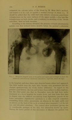
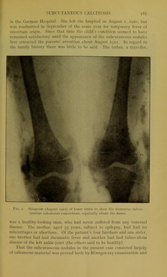
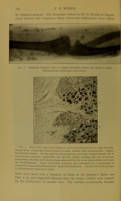
![[Frid st 8] DERMATOLOGY AND SYPHILOGRAPHY INDEPENDENT PAPER SUBCUTANEOUS CALCINOSIS OR MULTIPLE CALCI- FICATION IN THE SUBCUTANEOUS TISSUE By F. PARKES WEBER, M.D., F.R.C.P., Senior Physician to the German Hospital, London The patient, Anna G., then aged 7 years, was admitted to the German Hospital on July 30, 1912,1 on account of the presence of a large number of hard nodules in the subcutaneous tissue of the extremities and the portions of the trunk adjoining the extremities. Most of the nodules were smaller than an average pea, but some of them, especially those on the buttocks and about the knees, were much larger, the larger nodules having apparently arisen by the coalescence of several smaller nodules. The face, head, thorax, and abdomen were practically free. On the child’s admission to the hospital the skin over one of the nodules was ulcerated, and the skin was inflamed and adherent over one or two others, but the nodules, as a rule, gave rise to no pain or tenderness, and seemed to have developed without the child being aware of their existence. The lymphatic glands in the groins and axillae, and some in the neck, were moderately enlarged. The liver and spleen could not be felt, and the child seemed to be free from any visceral disease. The urine contained no albumin or sugar. There was no fever. Brachial systolic blood-pressure, no mm. Hg. Ophthalmoscopic examination (right eye) showed nothing abnormal. Blood examination (October 8, 1912) : Haemoglobin, 70 per cent. ; red cells, 4,070,000 per cubic milli- metre of blood; colour index, 0-9; white cells (after a meal). 11,200 per cubic millimetre of blood. The differential count of the white cells gave : Polymorphonuclear neutrophiles, 65 per cent.; small lymphocytes, 27 per cent. ; large lymphocytes, 2 per cent. ; large mononuclears, 3 per cent. ; transitionals, 2 per cent. ; eosinophiles, 1 per cent. ; mast- , cells, none in the count. The red blood-corpuscles appeared normal. The coagulation time (estimated by Sir A. E. Wright’s tubes at 250 C.) was about three minutes. On October 7, 1912, Dr. G. R. Ward kindly 1 The case was shown and described at the Clinical Section of the Royal Society of Medicine on October 11, 1912 (Proceedings, 1913, vi, p. 14). See also F. P. Weber, British Journal of Children’s Diseases, London, 1913, x, p. 97. XIII N 2](https://iiif.wellcomecollection.org/image/b22445110_0005.jp2/full/800%2C/0/default.jpg)