Abstract of lectures on clinical medicine : delivered in the Royal Infirmary of Aberdeen during session 1866-67 / by James W.F. Smith and Archibald Reith.
- Smith, James W. F.
- Date:
- 1867
Licence: Public Domain Mark
Credit: Abstract of lectures on clinical medicine : delivered in the Royal Infirmary of Aberdeen during session 1866-67 / by James W.F. Smith and Archibald Reith. Source: Wellcome Collection.
25/68 (page 25)
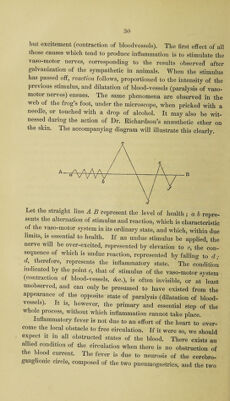
![[This patient continued to improve until the 26th March, when she w*as suddenly seized with pain in the left side and shortness of breathing, and died next day. There was no return of the hoemoptysis. At the autopsy on the 29th a considerable serous effusion was found in the left pleural cavity. The lower lobe of the left lung was carnified, and contained the remains of several nodules of pulmonary apoplexy. Heart much enlarged. Both auricles considerably dilated, and their walls thinned. Mitral orifice constricted to one-third of its natural size. Aortic valves incom¬ petent, with fibrous vegetations on their free border.] LECTURE—18th March, 1867. PHTHISIS-SUDDEN DEATH—GENERAL REMARKS ON DYSPEPSIA. A patient in St. Luke’s Ward, suffering from phthisis in the apex of the right lung, in the third stage, but whose general health and strength was not much reduced, and who was feeling unusually well one evening, died suddenly the following morning. He awoke about two in the morning, said he had had a good sleep and felt very comfortable, and in an hour after he was observed by some of the patients to be gasping for breath, and was dead before the night nurse could be got. From the nature of the case and the history, it seemed probable that death had been caused by embolism or some obstruction in the pulmonary artery. At the autopsy the adipose tissue, both sub¬ cutaneous and in the cavities, was found to be abundant. Both lungs contained tubercle and caverns in their apices. The heart weighed 13 oz., its cavities large, dilated, the walls not thickened, pale and flabby; all the cavities filled with coagula, which in the right ventricle were colourless. The large vessels similarly filled, especially the pulmonary artery, which in its trunk and all its large branches contained a colourless continuous fibrinous coagulum. Kidneys large, flabby, and fatty. Other organs healthy. The cause of death was thus found to be the formation of coagula in the right ventricle and pulmonary artery, consequent on failure of action on the part of the dilated and enfeebled heart. I)](https://iiif.wellcomecollection.org/image/b30567853_0025.jp2/full/800%2C/0/default.jpg)