Licence: Public Domain Mark
Credit: A text-book of histology / by Frederick R. Bailey. Source: Wellcome Collection.
Provider: This material has been provided by the Augustus C. Long Health Sciences Library at Columbia University and Columbia University Libraries/Information Services, through the Medical Heritage Library. The original may be consulted at the the Augustus C. Long Health Sciences Library at Columbia University and Columbia University.
130/674 page 106
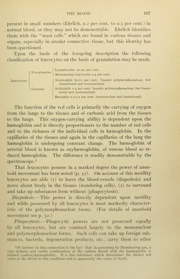
![down of the connecting cords, leaving several separate nuclei or nuclear segments. Granules in small numbers may be present in the protoplasm of any of the leucocytes, but the protoplasm of about 70 per cent, of all leucocytes is so distinctly and regularly granular that by some authors a prim.ary division into granular leucocytes and non-granular leuco- cytes is made. Under this classification, lymphocytes and some mononuclear leucocytes are placed in the non-granular group, while transitional leucocytes, polym.orphonuclear leucocytes and some mononuclear leucocytes, are placed in the granular group. Aniline dyes may be divided into acid, basic and neutral, according to whether the coloring matter is an acid, a base, or a combination of an acid and a base, and the granules of the granular leucocytes react in a definite manner to these dyes, thus allowing the following classification: f I neutrophile. Granular leucocytes] 2 acidophile (eosinophile). [ 3 basophile. As the neutrophile granules are fine and the eosinophile granules coarse, a classification of leucocytes into finely granular and coarsely granular is sometimes made.' 1. Neutrophile Leucocytes.—These are the most numerous of all leucocytes, making up about 68 per cent. Their protoplasm is thickly studded with very fine granules which stain violet with a mixture'of eosin (acid) and toluidin blue (basic). Most neutrophiles are polymorphonuclear, a few are transitional. They have a wide distribution, being found not only in the blood itself, but in the spleen and lymph nodes and as wandering cells in various tissues and organs. 2. Acidophile Leucocytesl or, because the most common acid dye used is eosin, eosinophile. The granules in these cells are coarse and sharply defined. They stain strongly with acid dyes. Eosino- philes are mainly polymorphonuclear, m.ore rarely they are transi- tional. They make up from i per cent, to 4 per cent, of all leuco- cytes. In certain pathological conditions their number is greatly increased. 3. Basophile Leucocytes.—The granules in these cells are rather coarse and irregular in shape and are distributed unevenly through the cytoplasm. They stain strongly with basic dyes. They are](https://iiif.wellcomecollection.org/image/b2122948x_0130.jp2/full/800%2C/0/default.jpg)
 No text description is available for this image
 No text description is available for this image
 No text description is available for this image