Licence: Public Domain Mark
Credit: A text-book of histology / by Frederick R. Bailey. Source: Wellcome Collection.
Provider: This material has been provided by the Augustus C. Long Health Sciences Library at Columbia University and Columbia University Libraries/Information Services, through the Medical Heritage Library. The original may be consulted at the the Augustus C. Long Health Sciences Library at Columbia University and Columbia University.
305/674 page 281
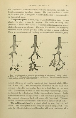
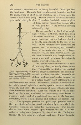
![beneath the epitheHum. From these, terminals pass to the secreting cells. It is probable that the saHvary glands also receive sympathetic fibres from cells of the superior cervical ganglia. TECHNIC (i) The salivar)^ glands should be fixed in Flemming's fluid (technic 7, p. 7), or in formalin-JNIiiller's fluid (technic 5, p. 7). Sections are cut as thin as possi- ble, stained with haematoxylin-eosin (technic i, p. 20), and mounted in balsam. (2) For the study of the secretory activities of the gland cells, glands from a fasting animal should first be examined and then compared with those of a gland the secretion of which has been stimulated by the subcutaneous injection of pilo- carpine. Fix in Flemming's or in Zenker's fluid (technic 9, p. 8). Examine some sections unstained and mounted in glycerin, others stained with hasma- toxylin-eosin and mounted in balsam. (3) The finer intercellular and intracellular secretory tubules are demon- strated by Golgi's method. Small pieces of absolutely fresh gland are placed for three days in osmium-bichromate solution (3-per-cent. potassium bichromate solution, 4 volumes; i-per-cent. osmic acid, i volume), and then transferred with- out washing to a o.7S-per-cent. aqueous solution of silver nitrate. Here they remain for from two to four days, the solution being frequently changed. The processes of dehydrating and embedding should be rapidly done, and sections mounted in glycerin, or, after clearing in xylol, in hard balsam. Pancreas The pancreas is a compound tubular gland. While in general similar to the salivary glands, it has a somewhat more complicated structure. A connective-tissue capsule surrounds the gland and gives off trabeculae which pass into the organ and divide it into lobules. In some of the lower animals, as for example the cat, these lobules are well defined, being completely separated from one another by connective tissue. In this respect they resemble the lobules of the pig's liver. A number of these primary lobules are grouped together and surrounded by connective tissue, which is considerably broader and looser in structure than that separating the primary lobules. These constitute a lobule group or secondary lobule. In the human j)ancreas the division into lobules and lobule groups is much less distinct, although it can usually be made out. This is due to the incompleteness of the connective-tissue septa, the human pancreas in this respect resembling the liuman liver. Rarely the human pancreas is distinctly l()])ulated. The gland has a main excretory duct, the pancreatic duct or dud of Wirsung. In many cases there is also a secondary excretory dud,](https://iiif.wellcomecollection.org/image/b2122948x_0305.jp2/full/800%2C/0/default.jpg)
 No text description is available for this image
 No text description is available for this image
 No text description is available for this image