Licence: In copyright
Credit: A manual of midwifery / by Alfred Lewis Galabin. Source: Wellcome Collection.
Provider: This material has been provided by the Royal College of Physicians of Edinburgh. The original may be consulted at the Royal College of Physicians of Edinburgh.
912/982 (page 882)
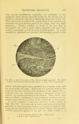
![manner which has been compared to the function of tlie syncytium in the development of the normal placenta. Malignant cells have been found in thrombi within the vessels. In the growing margin, where the growth is infiltrating the muscular wall, cells or groups of cells appear to have a special tendenc}'^ to penetrate the venous sinuses and engraft themselves on the interior of their walls. In this way is accounted for the very rapid formation of metastatic deposits, which occur generally through the vessels and not through the 13'inphatics, and ospeciall}'^ in the lungs, 'riiere is a marked tendency to necrosis, and, in the typical part of the tumour, there are no vessels among either cells or syncytium. In the infecting margin, however, small groups of cells, or small masses of syncj’tium, even as small as single cells, appear to be springing up in the stroma, so that this part of the growth may ap])roximate in character to the third variety, next to be described. (8) In comparatively rare cases, generally of an advanced kind, where the jiatient has died from the disease without operation, there is found no considerable development or branching processes of syncytium, but only comparatively small masses of nucleated ])rotoplasm, combined with a large })roportion of discrete cells, 'riiis kind of growth was at first regarded by many authorities as sarcoma. Hut it is now generally held that the small protoplasmic masses, whicli have tlie staining (pialities of s^meytium, really are syncytium, and that this variety is develo])ed out of the second, the more typical portions of the growtli having become necrosed. If the syncytium of tlie villi in retained placenta after abortion (Figs. I fi7, 108, ]). .‘19.0), and in vesicular mole (Figs. 172, 173, p. 402), is compared with f'igs. 328, 329, there apiJears to be a gradation toward the structure seen in the so-called decidnoma malignum ; and an intermediate stage between Figs. 172, 173, and ]<’igs. 328, 329, is furnished by cases in which chorionic villi are ])resent in the malignant growth, such as that figured by Haidtain. The author met with one case, before decidnoma malignum had attracted attention, in which a w’oman, near the usual time of the menopause, had a vesicular mole. This was followed within a few' weeks by an intra-nterine growth, which bled freely, and dis- charged gelatinous masses vagimmn. Sections of the grow'th had the structure of myxoma, and no chorionic villi, degenerated or otherwise, w'ere present. For some months the growth appeared to be running a malignant course, but eventually, after repeated clearing out and curetting the uterus, it died out, the meno2)ause became established, and the patient has remained over ten j’ears free from recurrence. This appeal's to have been probably an implantation of mj'xoma from the stroma of the degenerated villi : and it is notable that it did not show the malignancy of deciduoma malignum. In a few cases deciduoma maligmun has been described as](https://iiif.wellcomecollection.org/image/b21932645_0912.jp2/full/800%2C/0/default.jpg)