On the arrangement of the fibres of the heart / by Henry Searle.
- Searle, Henry.
- Date:
- 1838
Licence: Public Domain Mark
Credit: On the arrangement of the fibres of the heart / by Henry Searle. Source: Wellcome Collection.
Provider: This material has been provided by The Royal College of Surgeons of England. The original may be consulted at The Royal College of Surgeons of England.
7/16
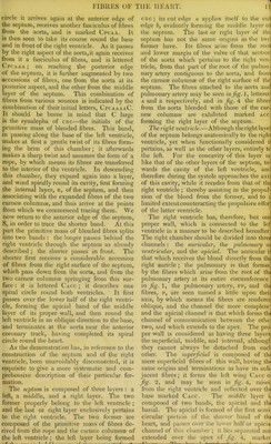
![coronary track, joins a band emerging from the septum, and thus forms the apicial half of the posterior boundary of this ventricle. It is raised from its situation, but when replaced its edge, which is everted by the probe, applies itself to the anterior boundary of this cavity. This layer cannot often be so extensively dis- connected from its superjacent bands as this figure represents. The thud stage of the dissection.—Having separated the layers composing the right or proper wall of the right ventricle, the next pro- ceeding consists in detaching and unwinding the band and layers composing the left ventri- cle. First, the detachment of the basial band. As this band has already been detached over the right ventricle in the second stage of the dissection, it is necessary to resume its separa- tion at the posterior coronary track. But as the further separation is somewhat difficult, it will be rendered less so if the remaining portion of this band be first examined in jig. 5, wherein it is represented detached. When in its natural situation it forms the uppermost third of this, the left ventricle, and its lower fibres overlap a part of those which occupy the middle third. The fibres which overlap the others in taking an oblique course towards the base reach the brim of the ventricle and pass over it, while the under fibres of this band are appearing in succession, and taking a similar spiral course until the whole bundle of fibres is twisted in the form of a rope. In order, there- fore,to trace out and detach this band as it becomes transformed into a rope, it is requisite to com- mence near the posterior coronary track (pet), in a continuous line with the lower edge of its former portion, introducing the handle of a scalpel obliquely upwards so as to detach the fibres which overlap those of the middle third, md to carry the separation so far up as will reach those marked a, coming obliquely down from the aorta. In conducting this separation from left to right it is soon found that the fibres af this bundle, instead of overlapping others, become themselves by twisting overlapped, rendering it necessary, therefore, to turn gra- dually the handle of the scalpel obliquely downwards, tracing the rope according to its windings. Two scalpels will be required in inducting the further separation. The next step should be preceded by viewing the fibres of the rope in fig. 3, descending md radiating into a layer which sweeps round the cavity of this ventricle. The heart should now be placed in a small cup or jar of a size that will support it with its base upwards, and -hen, with the scalpels employed vertically, the separation should be proceeded with, and in massing through the septum a vertical section ihould be made through the aorta in the ine of separation, which should be pursued onnd anr) round, and nrnpressi veliL_dfi£n£^- of the heart, and which now pass over tire scal- pels, should be divided; the incision being made along the side of the posterior edge oi the septum. A section should be made through the rope also, which allows the right ventricle to be raised from the left, and the heart to be unwound as far as the separation has been car- ried. There yet remains a mass of fibres around the cavity of the left ventricle to be de- tached. This last process of separation shoulc be conducted in a contrary direction to tha which has hitherto been adopted, viz. from right to left, until the internal membranous lining is exposed, and which should be torn in order tc lay open this chamber. The heart can now be unwound and extended as in fig. 1, placing the left ventricle, Iv, ati one end and the right at the other, removing that section of the aorta, aa, connected to tha right ventricle from its counterpart which ex-1 clusively pertains to the left, and which is hid-] den by the rope, r r ; removing also the two! portions of the bisected rope to the two most] distant diagonal points in this view. Tha niche, Cpc, indicates the part occupied by the divided band which passed along the mid- dle third of the heart. The second method of demonstration.—Thpj formation, or winding up of the fibres* of the heart. This description comprehends! the retracing of the fibres from the centre to the circumference, showing their respective origins, associations, courses, connexions, and terminations, also the manner in which they are wound up to form the two ventricles into one compact conical body. The first stage consists in retracing the su- perficial layer from its origins to its termina- tions. It is necessary to commence at the very centre of the heart—the interior of the left ventricle, whence spring the fibres composing its main bulk. Fig. 4, at its right extremity, exhibits the left ventricle, Iv, laid open, exposing the two carneae columnae, cc and cc, one of which is placed out of its situation, in order tOj show the interior of the chamber. The fibres of the two carneae columnae, cc and cc, ex- pand in a fan-like manner; those of the rope, rr, expand in a similar manner; the radiated fibres of each of these three bodies wind round the axis of this ventricle forming its parietes; and as they wind so as to form an inverted cone, it is clear that the inmost fibres alone can reach the apex. Accordingly, a fasciculus of the inmost fibres from each of these three bodies, marked c, r, and c respectively, pass down to the apex associated together, and in their course make a gentle twist from left to right, gradually contracting the cavity to a point and closing it; they then twist sharply round upon each other and complete the apex marked crc conjointly, so that by means of tins twistimr the internal fibres are .-PnrW.L](https://iiif.wellcomecollection.org/image/b22386476_0009.jp2/full/800%2C/0/default.jpg)