Licence: Public Domain Mark
Credit: Structure and functions of the brain and spinal cord. Source: Wellcome Collection.
Provider: This material has been provided by the Augustus C. Long Health Sciences Library at Columbia University and Columbia University Libraries/Information Services, through the Medical Heritage Library. The original may be consulted at the the Augustus C. Long Health Sciences Library at Columbia University and Columbia University.
122/274 page 108
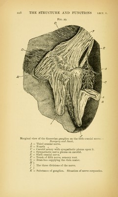
![Fig. 29. J- Marginal view of the Gasserian ganglion on the fifth cranial nerve.— Bov.rgery and Jacob. A = Third cranial nerve. B = Fourth „ C = Carotid arterr with sympathetic plexus upon it. iS = Sympathetic nerve plexus on carotid. J^ = Sixth cranial nerve. -F = Trunk of fifth nerve, sensoiy root. G — Branches supplying the dura mater. S] I - The three divisions of the ner^e. •/ ) K = Substance of ganglion. Situation of nerve corpuscles.](https://iiif.wellcomecollection.org/image/b21218262_0122.jp2/full/800%2C/0/default.jpg)