Licence: Public Domain Mark
Credit: Structure and functions of the brain and spinal cord. Source: Wellcome Collection.
Provider: This material has been provided by the Augustus C. Long Health Sciences Library at Columbia University and Columbia University Libraries/Information Services, through the Medical Heritage Library. The original may be consulted at the the Augustus C. Long Health Sciences Library at Columbia University and Columbia University.
204/274 page 190
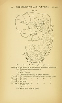
![Fig. 44. Human embryo.—His. Showing the peripheral nerves. Ill to XII = The crania] nerves in order from the third to the twelfth. Cb — Developing cerebral hemispheres. m = Mid brain. 4 = Fourth ventricle. b — Commencement of bulb, or meduUa oblongata. C(i to 8) — The cervical nerves and ganglia on their posterior roots. Z> (i to 12) = The dorsal „ „ ,, ,, L (i to 5) = The lumbar „ ,, „ ,, ^S'(i to 5) = The sacral „ ,, „ „ Co {i to 2) = The coccygeal „ „ „ „ Ht — Ventricle of heart. i — Intestine. is = Sciatic nerve cut at its origin.](https://iiif.wellcomecollection.org/image/b21218262_0204.jp2/full/800%2C/0/default.jpg)