Licence: Public Domain Mark
Credit: Structure and functions of the brain and spinal cord. Source: Wellcome Collection.
Provider: This material has been provided by the Augustus C. Long Health Sciences Library at Columbia University and Columbia University Libraries/Information Services, through the Medical Heritage Library. The original may be consulted at the the Augustus C. Long Health Sciences Library at Columbia University and Columbia University.
69/274 page 55
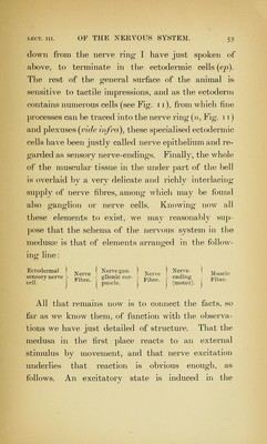
![down from the nerve ring I have just spoken of above, to terminate in the ectodermic cells (ep). The rest of the general surface of the animal is sensitive to tactile impressions, and as the ectoderm contains numerous cells (see Fig. 11), from which fine processes can be traced into the nerve ring {n, Fig. 11) and plexuses {vide infra), these specialised ectodermic cells have been justly called nerve epithelium and re- garded as sensory nerve-endings. Finally, the whole of the muscular tissue in the under part of the bell is overlaid by a very delicate and richly interlacing supply of nerve fibres, among which may be found also ganglion or nerve cells. Knowing now all these elements to exist, we mav reasonably sup- pose that the schema of the nervous system in the medusae is that of elements arranged in the foliow- ino- line: o Ectodermal ] ( Nerve gan- ] | Nerve- ) j,^^^,^ sen,orv nerve , ^„„ ghoo.c cor- ,,„,^ endrng^ ,.;,,,. All that remains now is to connect the facts, so far as we know them, of function with the observa- tions we have just detailed of structure. That the medusa in the first place reacts to an external stimulus by movement, and that nerve excitation underlies that reaction is obvious enough, as follows. An excitatorv state is induced in the](https://iiif.wellcomecollection.org/image/b21218262_0069.jp2/full/800%2C/0/default.jpg)