Surgical anatomy : a treatise on human anatomy in its application to the practice of medicine and surgery / by John B. Deaver.
- Deaver, John B. (John Blair), 1855-1931.
- Date:
- 1899-1903
Licence: In copyright
Credit: Surgical anatomy : a treatise on human anatomy in its application to the practice of medicine and surgery / by John B. Deaver. Source: Wellcome Collection.
34/718 (page 32)
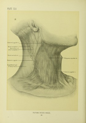
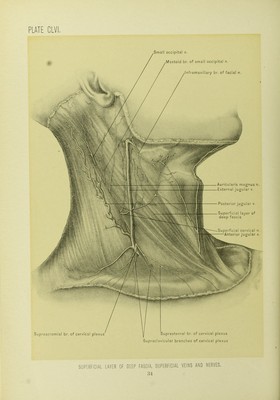
![descending from the submaxillary region to the upper part of the chest-wall, where pointing may occur. Dissection.—The platysma should now be removed, cutting it across near the clavicle and reflecting it upward to its insertion into the jaw, thus exposing the subcutaneous portions of the superficial branches of the cervical plexus of nerves, the infra-maxillary branch of the cervico-facial division of the facial nerve, and the anterior, external, and posterior jugular veins. The external jugular vein arises in the substance of the parotid gland, and is formed l)y the union of the posterior auricular vein and the posterior division of the temporo-maxillary vein. It runs down the neck in a line drawn from the angle of the lower jaw to the middle of the clavicle, first passing over the sterno- mastoid muscle, and then along its posterior border to the root of the neck, there piercing the superficial layer of the deep cervical fascia to enter the subclavian vein in the subclavian triangle. This fascia is so closely attached to the vein that at the root of the neck, if the vein be divided, it will remain open. The auricu- laris magnus nerve, a branch of the cervical plexus, accompanies the vein in its vipj)er part, and the superficial cervical l)rancli of the same plexus passes beneath it at about the middle of the course of the vein. The posterior external jugular, transversalis colli, and supra-scapular veins empty into the external jugular vein. Near the angle of the lower jaw the external jugular communicates with the internal jugular vein by a lai'ge branch, farther down with the anterior jugular-,- and, at times, with the cephalic vein by a brancli (jugulo-cephalic) which passes over the clavicle. The anterior jugular vein occasionally empties into the external jugular instead of into the subclavian vein. The external jugular vein contains a ])air of valves at its point of entrance into the subclavian, and another pair about one inch or one and one-half inches above this point ; these valves can not prevent the reflux of blood into tlie external jugular vein, and in certain cardiac and aortic diseases, especially in tricuspid insufficiency, a pulsation in the external jugular vein synchronous with the cardiac systole may be observed. The portion of the vein between the valves is dilated ; tliis poi'tion is called the sinus. The external jugular vein varies in size—when the anterior and posterior jugular veins are large, the external jugular vein is small, and vice versri. In some instances two external jugular veins may l)e observed upon each side of the neck. The superficial cervical nerve may, at times, l)e seen to pierce the wall of the vein. Venesection.—The operation of ])hlebotomy, or venesection, may be per- foi-med upon the external jugular vein. AVhen the lower portion of the vein is selected for the operation, the direction the fibers of the platysma myoidcs muscle should be borne in mind, and the incision be made across them. They will then retract and pull the wound open, tlius allowing the blood to flow freely and](https://iiif.wellcomecollection.org/image/b20415345_0034.jp2/full/800%2C/0/default.jpg)