The pocket Gray, or, Anatomist's vade-mecum / by the late Edward Cotterell.
- Cotterell, Edward, 1857-1898.
- Date:
- 1901
Licence: In copyright
Credit: The pocket Gray, or, Anatomist's vade-mecum / by the late Edward Cotterell. Source: Wellcome Collection.
33/284
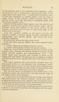
![The tterygo-maxillary ligament extends from the hamular process to the posterior edge of the mvlo-iiyoid ridge of rri*erior maxilla. It separates buccinator from superior con- strictor of pharynx, giving origin to both. Risorius : fascia covering masseter(F)—apex of depressor anguli oris(F) (Facial). [Draws angle of mouth back.] (This muscle is part of the platysma of neck.) TEMPORO-MAXILLARY REGION. Masseter: Superficial part. Malar process of superior maxilla. Anterior % of lower border oT zygoma(TA)—angle and lower ot outer surface ot ramus(F). Dccjp part. Posterior § lower border ana inner surtace of zygoma(F)—upper J of ramus and outer surface of coronoid process(F) (Inferior maxillary). [Muscle of mastication; elevates lower 1 aw and draws it The niassetericjascia, a continuation of the deep cervical fascia, is attached above to the zygoma; continuing back- wards it invests parotid gland (parotid fascia), from the deep surface ot which the stylo-maxillary ligament proceeds. Temporal: temporal fascia and fossa(F)—internal surface and forepart ot coronoid process ot inferior maxilla as tar as last molariat) (interior maxillary), fMuscle of mastication, closingjjjmjth; antenornbre^rotrude lajvwpostenorjetract.] The temporal fascia is attached above to the temporal ridge, and divides below into two layers, which are attached to inner and outer edges of superior border of zygoma. It covers the temporal muscle, and between the two layers are the temporal branch of temporo-malar nerve, and the orbital branch of superficial temporal artery. PTERYGO-MAXILLARY REGION. External pterygoid: pterygoid ridge and surface below on great wing ot sptienoid. outer surtace ot external pterygoid plate(F)—pterygoid depression in front of neck ot inferior maxilla and mte^articularti Pro-cartilageol temporo-maxillTTry jointO) (Interior maxillary). [Muscle, ot mastication ; both acting together protrude lower jaw : acting alternately cause grinding movements, each moving; jaw to opposite side.] Between sbhenoidal and pterygoid attachments. the internal maxillary anery dips down to reach spheno-maxillarv fossa, and the buccal ana anterior deep temporal nerves appear. Internal pterygoid : inner surface of external pterygoid plate, tuberosity of palate bone, and tuberosity of superior](https://iiif.wellcomecollection.org/image/b28079838_0033.jp2/full/800%2C/0/default.jpg)
 No text description is available for this image
 No text description is available for this image
 No text description is available for this image