The pocket Gray, or, Anatomist's vade-mecum / by the late Edward Cotterell.
- Cotterell, Edward, 1857-1898.
- Date:
- 1901
Licence: In copyright
Credit: The pocket Gray, or, Anatomist's vade-mecum / by the late Edward Cotterell. Source: Wellcome Collection.
54/284
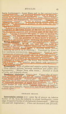
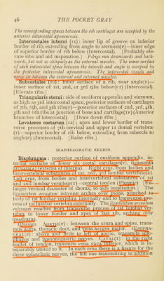
![The corresponding spaces between the rib cartilages are occupied by the anterior intercostal aponeurosis. Intercostales interni (u): inner lip of groove on inferior border of rib, extending from angle to sternum(F)—inner edge of superior border of rib below (Intercostal). [Probably ele- vate ribs and aid inspiration.] Fibres run downwards and back- wards, but not so obliquely as the external muscles. The inner surface of each intercostal space between the tubercle and angle is occupied by the ■ posterior intercostal aponeurosis. The intercostal vessels and nerve lie between the external and internal muscles. iaubcostaies (io) : inner surtace ot a rib, near angle(F)— inner surface of ist, 2nd, or 3rd rjbs below(FT) (Intercostal). [Elevate ribs.] Triangularis sterni: side of ensiform appendix and sternum, as high as 3rd intercostal space, posterior surfaces of cartilages of 7th, 6(h, and 5th ribs(F)—posterior surfaces of 2nd, 3rd, 4th, 5th and Cth ribs at junction of bone and cartilage(TF) (Anterior branches of intercostal). [Draw down ribs.] Levatores costarum (12) : apex and lower border of trans- verse processes of 7th cervical and upper 11 dorsal vertebrae (t)—superior border of rib below, extending from tubercle to angle(F) (Intercostal). [Raise ribs.] DIAPHRAGMATIC REGION. Diaphragma : posterior surface of ensiform appendix, in- ternal surfaces ot lower six costal cartilages(F), ligamenTa arcuaT^A) (externamrnterna). kight crus, from bodies and Tm^rwertebrTT^^is!?mce^^r ist. 2nd*^^'*^imb<^Ter^eE^i^:). 'Lett crus, from bodies and mterverteprai suostances of ist and 2nd lumbar vertebrae(x) —central tendon (Phrenic). [En- larges vertical diameter of thorax, so aids inspiration.] The ligyuncntumarcuatini^^dennur^^c^Gsove^D^^s, cUtnehed to boclv'oi ist lumHar vertebra internally and to transverse pro- cessof ist lunibaTvert^jra externally. The hgamentum arcuatum extern//>» reaches trom transverse process_aLL£L nni ver- tebra to lower border and apex of last rib, arching over quadndus. * (jfenuu's. i\0R^nc(F) : between the cruraand_spine, trans- mi tsliarta. thoracic duct, ana vena azygos maior. CEsopha- geal(f) : aboveanrTittle to lett ot aortic, transmits cpso- 52^us and pneumogast^^^^^e^t^WAL(T) rTntJmjd^nt leaflet of tendon, transmits vena_cava55Euor. which is in- separably united to it. in eacfi crus there 1^ a fissure for the three splanchnic nerves, the left one transmitting in addition](https://iiif.wellcomecollection.org/image/b28079838_0054.jp2/full/800%2C/0/default.jpg)
 No text description is available for this image
 No text description is available for this image
 No text description is available for this image