Elementary anatomy and physiology : for colleges, academies, and other schools / by Edward Hitchcock and Edward Hitchcock Jr.
- Edward Hitchcock
- Date:
- 1861
Licence: Public Domain Mark
Credit: Elementary anatomy and physiology : for colleges, academies, and other schools / by Edward Hitchcock and Edward Hitchcock Jr. Source: Wellcome Collection.
Provider: This material has been provided by the National Library of Medicine (U.S.), through the Medical Heritage Library. The original may be consulted at the National Library of Medicine (U.S.)
29/452
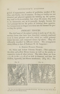
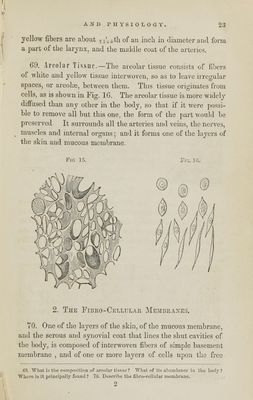
![yellow fibers are about T5'6()th of an inch in diameter and form a part of the larynx, and the middle coat of the arteries. 69. Areolar Tissue—The areolar tissue consists of fibers of white and yellow tissue interwoven, so as to leave irregular spaces, or areolae, between them. This tissue originates from cells, as is shown in Fig. 16. The areolar tissue is more widely diffused than any other in the body, so that if it were possi- ble to remove all but this one, the form of the part would be preserved. It surrounds all the arteries and veins, the nerves, muscles and internal organs; and it forms one of the layers of the skin and mucous membrane. Fig 15. Fro. ] G. 2. The Fibro-Cellular Membranes. TO. One of the layers of the skin, of the mucous membrane, and the serous and synovial coat that lines the shut cavities of the body, is composed of interwoven fibers of simple basement membrane , and of one or more layers of cells upon the free CO. What is the composition of areolar tissue ? What of Its abundance in tho body? Where is it principally found? 70. Describe the fibro-cellular membrane. 2](https://iiif.wellcomecollection.org/image/b21128674_0029.jp2/full/800%2C/0/default.jpg)