Different species of trypanosomata observed in bovines in India / by A. Lingard.
- Alfred Lingard
- Date:
- 1907
Licence: In copyright
Credit: Different species of trypanosomata observed in bovines in India / by A. Lingard. Source: Wellcome Collection.
Provider: This material has been provided by The Royal College of Surgeons of England. The original may be consulted at The Royal College of Surgeons of England.
37/58 (page 31)
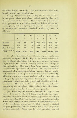
![the whole length anteriorly. Its measurements were, total length, SS'SCf* and breadth 5'4m- A single trypanosoma (PL II, Fig. ] 6) was also observed in tile spleen, whose protoplasm stained entirely blue, with the exception of the nuclei. This is particularly mentioned as it presented four nutritive nuclei, one dislocated, but only one blepharoplast undergoing division, Tlie measurements of twenty-one parasites described under (o) were as follows :— I II III IV V VI VII F Maximum 4-92 9-84 2-46 29 52 1312 29-86 3-28 Minimum 3-28 8-20 2-46 1804 3-28 35-26 2-46 Mean of 21 tryp. 3-98 8-86 2-31 22-63 8-32 46-10 3-25 Percentage of mean 8-63 19-22 5 01 49 10 1804 100-00 6 54 (c) A few involution forms of female trypanosomata were observed, as figured (PL II, Fig. 2), and described above in the peripheral circulation, but these were shorter, maximum length 28'60a'. the breadth varying from Pis anteriorly to 2‘Os posteriorly. The shape these forms assume, resembled somewhat the appearance of scimitar. The blepharoplast in one parasite had undergone division into two distinct nuclei, and occupied a clear space near to the posterior extremity, while the larger and compact nucleus, oval in form and 2'Os in length, lying in the long axis of the organism, occupied a position at the junction of the posterior third with the anterior two-thirds. The rest of the body of the parasite was occupied by the endoplasm, which stained a faint blue colour, and contained a thickly set mass of micro-granules. [d) These long or attenuated forms (PL II, Figs. 8—11) observed by mo for the first time, lying free in the paren- chyma of the spleen, presented a weird appearance, resembling snakes, threading their way through the corpuscles and cells. I was able to trace in a few instances the gradual suppression of the undulating membranes by their respective stained margins, proceeding from the smaller nuclei for a very short distance anteriorly. They were, however, but faintly marked](https://iiif.wellcomecollection.org/image/b22463525_0041.jp2/full/800%2C/0/default.jpg)